* Consultations * Laminitis/Founder * Thin soles * Navicular * Crushed/Low Heels * Foal development * Club feet * High Low Syndrome * Angular limb deformities
Pages
▼
Monday, January 12, 2015
Introduction to the mechanics of the lower limb and evaluation radiographically and clinically
Introduction to the mechanics of the lower limb and
evaluation radiographically and clinically
Sammy L. Pittman,DVM
Innovative Equine Podiatry and Veterinary Services,
Pllc
Considering a large component of lameness occurs in the
lower limb and the equine hoof a thorough understanding of the forces at play
are very helpful. We often examine and
treat lameness from a medical standpoint but are not fully recognizing and
changing the biomechanical properties that are very likely involved in creating
the lameness.
The detailed anatomy is covered at length in many text,
conversely, I want to focus on the functional anatomy as it relates to the
mechanical properties of the equine digit.
Consider the deep digital flexor tendon arising from the combined flexor
muscle bellies coursing distally over the palmar/plantar aspect of the fetlock
and pastern then over the navicular bone to attach to the semi-lunar crest on the solar aspect of the coffin bone. The
tendon attaches firmly to the bone and the bone is attached to the hoof wall
via the lamellar network. Think of these
combined anatomical structures as creating a sling or hammock for the boney
column. See figure 1 for a drawing
emphasizing the suspension and support components. Also consider the frog, ungual cartilages and
digital cushion as support structures accepting load that is determined by the balance of load from
the suspension system.
To further define the deep digital flexor tendon
suspension theory, consider a deep flexor contracture case versus a tendon
laxity case in young foals. The
contracture case has no load on the heels as they are suspended in the air via
the shortened tendon unit. Compare to
the tendon laxity case in which the toe is popping up and the heels and bulbs
are the weight bearing component. This
is a high suspension versus low suspension comparison and further describes how
the deep flexor tendon has a great influence on what structures are loaded
within the hoof capsule.
Figure 1 Suspension components and
support components
Now let's think about what load does to the hoof. For example compress one side of your
fingernail and watch it turn pale in color.
This is a load induced vascular compression that prevents the vascular
network from filling. The same goes for
the equine digit. When weight is placed
on the limb the vascular network is loaded and blood moves out of the loaded
areas to unloaded areas. This is easily
confirmed by performing venograms. As
long as the compression is temporary and balanced throughout the hoof it is of
no consequence. However when long term
compression occurs, bone and soft tissue suffer the effects of decreased
nutrient flow. This is evidenced by lack
of growth of sole and/or hoof wall and boney remodeling of the coffin
bone. Consider a high grade club foot
versus a crushed heel foot. Club feet
have trouble growing sole directly under the apex of the coffin bone and dorsal
hoof wall. Hooves with tendencies to
have long toes and low heels with difficulty growing heel. These are both load induced vascular
compressions secondary the loads determined by the deep flexor tendon
suspension. Figure 2 compares a foot with a severe negative palmar angle on the
left to a grade 3 club on the right. The
foot on the left has vascular compression under the wings of the coffin bone
and the foot on the right has compression under the apex of the coffin bone. The tighter suspension unit of the club
syndrome transmits a greater proportion of the load to the toe. The crushed
heel with less deep flexor suspension allowing more load at the heels.
Figure 2 Negative palmar angle
venogram on the left compared to a grade 3 club foot venogram on right.
Radiographic investigation with properly taken podiatry
style radiographs will allow definition of the areas that are chronically
loaded. Coffin bone shapes tell us the history of the loads that have been
applied to it. Wolfe's law describes
that bone remodels along lines of tension and compression Coffin bones shapes of club feet have a
characteristic bump about halfway down the face of the coffin bone, lipping at
the apex and resorption directly under the apex, secondary to forces acting
upon these regions from shortened musclotendinous unit. The articulation will also develop with more
dorsal orientation. Compare to the low
heel foot which will have a straight face and tip of the coffin bone with a scallop resorbed in the wings from the load
placed in this region. The articulation develops
further palmarly closer to the wings.
Evaluation of the center of rotation of the coffin joint will show that
the more upright clubby type foot has much less coffin bone dorsally when
compared to a lower heeled, long toe foot.
This effects the lever arm working against the deep flexor tendon that
is necessary to consider when treating the long toe low heel horse.
Taking consistent radiographs
before and after shoeing on all my foot lameness cases allows a greater
understanding of the mechanical properties that matter to the horse. Below is a review of the soft tissue
parameters that I routinely monitor.
Figure 3 Soft tissue parameters
Coronary
band to extensor process (CE) is measured from top of paste which is
applied at most proximal aspect hoof wall at the point of the last hair
follicle down to the extensor process of the coffin bone. This will range
from 8 to 30 mm in most healthy hooves. This number does not give you
much information as a single measured parameter. However, when monitored
and compared in serial radiographs, especially when monitoring an acute
laminitis case, it is extremely valuable. For example, an acutely laminitic
patient that measures 8mm on day 1 of clinical signs and then measures 18mm on
day 4. This is a 10mm distal displacement which is usually
accompanied by a 10mm decrease in sole depth as well and varying degrees of
rotational displacement.
Horn-Lamellar zone
(HL) is measured in two areas,
one proximal just below extensor process and one distal just above apex of
coffin bone. This will most commonly measure 15 mm in most light breed
horses but can be as high as 20mm in larger breeds, mules and donkeys.
This measure is expressed as proximal HL/Distal HL (15/15). Instead
of measuring only rotation this will give you a measurable displacement that is
more definitive than a generic rotation. Evaluating the dermal-epidermal
junction is also of great importance as it should split the horn
lamellar zone further defining each. This allows more specific
interpretation of changes in the HL zone. For example with laminitis the
L component of the HL zone will change not the H component. Early in
laminitis this may be the only notable change and an increase of 3-4 mm is a
significant finding and may have no measurable rotation. Several
important disease processes can be discovered in this zone and many foot
diseases such as clubs, chronic/acute laminitis, white line disease, keratomas
and abscesses have very unique qualities that can be shown here.
Sole Depth (SD) is measured from the tip of the coffin
bone down to most distal aspect of the sole. The cup is also of
importance as it is present to different degrees depending on health or pathology
and can also be falsely created with a hoof knife. This measurement is
expressed as SD/Cup. Healthy feet with no pathology will most commonly
carry 15mm of sole and a 2-3mm cup (15/3). This should be of upmost concern of
the vet/farrier team when striving to obtain soundness and health of the foot.
This should be the measurement at the day of the farrier visit.
Often thin soled horses are at 6-7mm of sole 8 weeks into the cycle and
this is a sign of a compromised foot that requires a different approach to
increase foot mass and health. Two measurements can be made to give you more
information, one at tip of coffin bone and one under wing of coffin bone.
Venogram findings suggest that a depth of 15mm is required to maintain a
healthy appearance to the solar vascular bed with robust and
correctly aligned terminal papillae
Digital Breakover
(DB) is measured from the tip of the coffin bone to where the foot or
shoe if shod would leave the ground. Healthy hooves that maintain
adequate SD and good digital alignment will commonly maintain a DB of 20-25mm.
Many times in perimeter fit shoes, depending on type of foot, bone angle,
and toe lever this number is considerably higher than ideal at the day of the
farrier visit and continues to lengthen throughout the cycle due to hoof
growth. This gives us a measurable lever arm that applies its force to the deep
digital flexor tendon and its subsequent force impacts on apex of the coffin
bone, dorsal hoof wall and navicular apparatus. Below I discuss toe lever
(TL) that in my opinion gives a more accurate understanding of the lever arm
involved.
Toe Lever (TL) can be expressed as static toe
lever or shod toe lever. Shod TL is measured from center of rotation of the
coffin joint to where the hoof/shoe would leave the ground and static TL is
measured from the center of rotation to the tip of the coffin bone. Shod TL we can effect and static we
cannot. Lower BA coffin bones
typically have a longer TL than higher degree. In my practice I see
static TL as short as 45mm to as long as 75mm in adult horses. Monitoring
this at a young age may allow us to apply orthotics that will decrease
the effective lever arm that antagonizes the lower limb.
Therapeutic shoe packages can be evaluated with regard to amount of lever
arm relief. Simply setting the shoe back only effects this measurement a
few millimeters and sometimes many lameness issues respond to a TL that is
3-4 times less than what is measured on their bare foot.
Bone Angle (BA) is the angle of the coffin
bone when viewed in a lateral radiograph. Average BA will be 50 degrees.
In my practice I have measured BA's as low as 36 degrees in very low
heeled and long toed horses to 70 degrees in club feet. The shape of the
coffin bone determines the shape of the hoof. Most of the time the horses
that have low heel long toe conformation will have a less than 50 degree bone
angle with a long measurable toe lever (see below) and the opposite is true for
upright club feet. Granted, horses that have overgrown unkempt feet may have
crushed heels and a long toe but may have a good BA. I feel that
monitoring this parameter early in life could potentially identify feet that
may have a common sequelae with regards to lameness later in life. For
example, a horse with a 42 degree BA and a 70mm Toe lever may be at higher risk
of hyperextension injuries of the pastern, coffin and fetlock joint and
increased tension strain on deep digital flexor tendon, and navicular apparatus
when compared to a coffin bone with a lower bone angle and shorter toe
lever. If we could identify this early in a horse's career and change the
shoeing protocol to better manage this handicap maybe we could reduce the
amount of wear and tear to some degree.
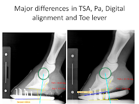
Tendon Surface Angle (TSA) is measured on this distal part of the navicular bone compared to a level ground marker. This is relative to the course of the deep digital flexor tendon takes at turns to attach to the coffin bone. Monitoring the change of TSA with your applied orthotic is of value especially cases that show navicular bone lesions in this region. Simply changing DB may be beneficial in many cases however raising PA and TSA is often required to be therapeutic.
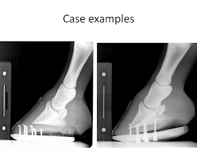
Figure 4 Pre and post shoeing
measurements
Figure 5 low versus high on a 6 mo old
foal
Figure 6 long toe low heel/neg pa hoof
versus a grade 3 club
Clinical evaluation is directed at evaluating growth
rings, hoof quality and length from the widest part of the foot forward. Clubby feet with higher deep flexor tension
will have growth rings that are narrow at the toe and get wider towards the
heel. Again this is secondary to the
loads creating a vascular compression and decreased nutrient flow to these
areas. This widest part of the foot
which correlates very close to the center of rotation will typically be in the
middle of or just in front of the middle of the hoof. The low heel type foot with lower suspension
properties within the deep flexor tendon will have more load in the heels. This results in growth rings that are wider
at the toe and narrower at the heel. The
widest part of the hoof is typically in the palmar third creating a long lever
arm.
Figure 7 Grade 2.5 club with heel
outgrowing toe versus a crushed heel with toe outgrowing heel
Note in
figure 7 the club foot on the has growth rings that diverge from toe to
heel and the crushed heel diverges from the heel to the toe.
Other aspects to consider from the solar view is frog characteristics. Typically with upright clubby feet that present as adults will have recessed atrophied frogs compared to the crushed heel hoof that will have a robust strong frog. Paying close attention to bulges will also further define regions of excessive load. This will occur around the apex of the frog with higher grade clubs and laminitic feet and just under the wings in negative palmar angle or crushed heel feet. Another good tool is watching your patients go in soft footing and watching what the coronary band, toe and heel does. Watch the heel and toe for sinking into the forgiving footing. Watch the coronary band. Does is stay level, rotate forward and more positive or backwards and more negative. This will also give you a good indication of the deep flexor system and what is will allow.
This
mechanical scenario has implications that must be considered in every foot disease. Using the podiatry style radiograph and
venogram to determine compromised areas and design a therapeutic shoeing
program is paramount to have repeatable success. Simply altering the toe lever length by
setting a shoe back, rolling or rockering the toe and use of natural balance
shoes has proven to offer mechanical advantage but has its limitations. Greater success is obtained by altering and
monitoring sole depth, palmar angle, tendon surface angle and digital
alignment. Difficult cases in my
practice have pre and post shoeing radiographs at every visit. The pre-shoe gives you information regarding
how the horse responded to your mechanical therapy with regards to palmar
angle, sole depth and digital alignment. The post shoeing radiographs sets a
new baseline and confirms you have accomplished your therapeutic goal with your
trim and shoe application.


