We had a great day resetting and rechecking many of the cases we used as demo's during Dr. Ric Redden's in depth equine podiatry lecture and demonstration. We had some return students and some new ones attending. I want to thank Clyde Brown and Animal Health Supply for allowing us to congregate at their place of business.
Below is several follow up images and short discussion of each case. Also look back at the previous blog entry for initial images and therapeutic shoe applied. October clinic images link
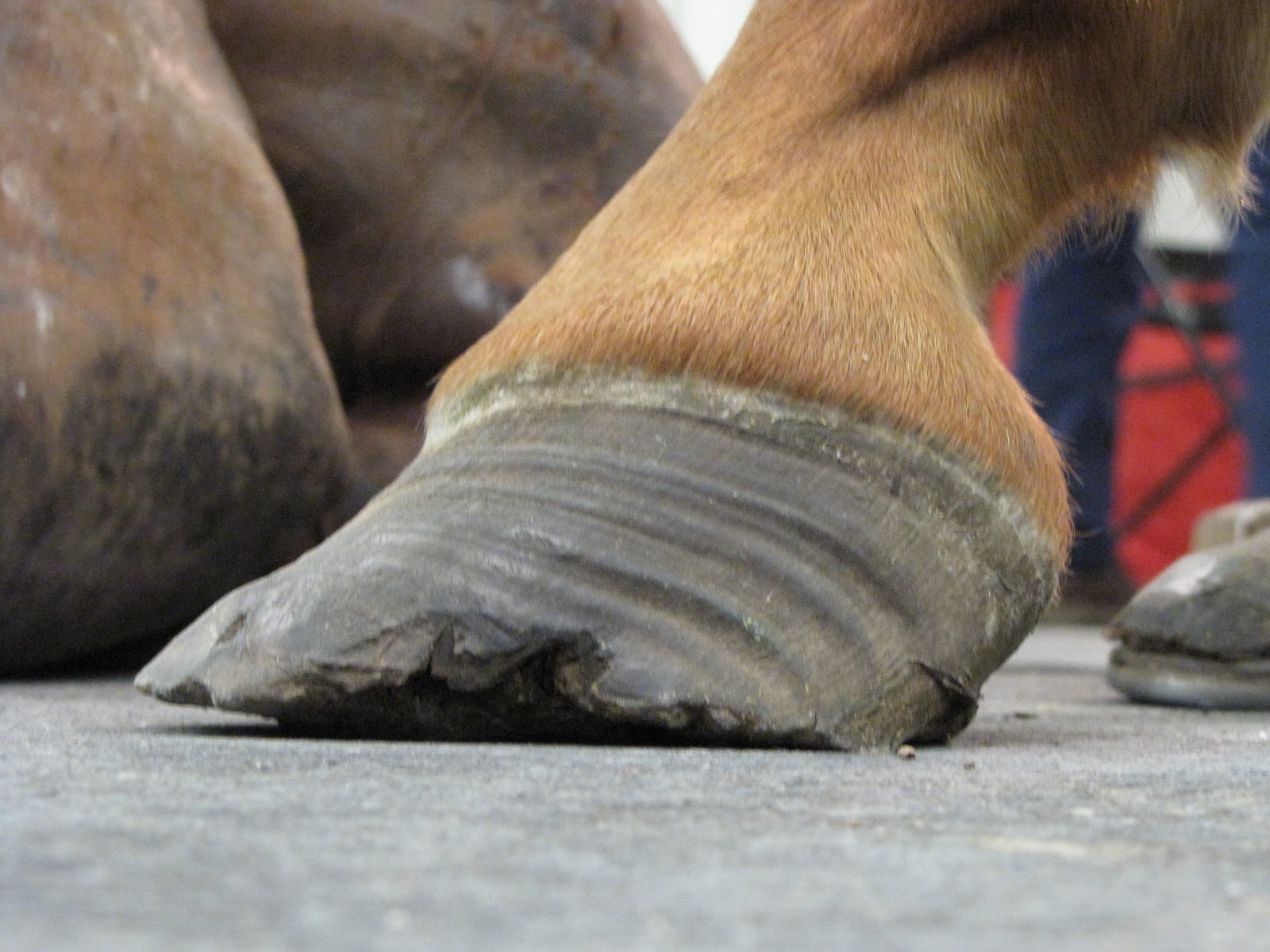
White line disease Case: Sole depth improved by 4mm but white line lesion failed to grow down at same rate and decision was made to remove hoof wall to expose oxygen and allow cleaning. Owner reports that he is running around like a youngster again and is more comfy than is has been in a long time.


Club foot case: This horse lost the rocker rail shoe applie to the foot opposite the grade 3 club (which is also a club) and regular farrier applied a flat steel keg shoe to keep foot protected. Note the horn lamellar zone divergence. One could call this rotation which would be non specific. The divergence is created by the club syndrome stretching to lower horn to bone attachments. This is confirmed by evaluating the dermal-epidermal junction and measuring the horn zone compared to the lamellar zone. If the lamellar zone was larger than the horn zone one could conclude a laminitis as this is lamellar swelling. In this case it is chronic stretching of the lamellar bone secondary to the constant pull of the deep digital flexor unit.
The Grade 3 club grew more sole in the rocker rail than did the lower grade club in a flat shoe. This information tells us that placing the tendon sling in freedom with the rocker shoe allows better nutrient and blood circulation through unloading of the sole via reduced deep flexor tension. We placed the grade 2 club (Left Front) in a rockered trim with rockered steel keg shoe to also place the tendon sling in release. We will be to see a more rapid sole mass recovery in this hoof as well at the next reset. Owner reports excellent comfort and has adjusted very well to the new shoeing approach.
Chronic Lamintis case: Farrier was a student and he reports horse is moving very nice. Horse was able to stand comfortably for each shoe reset. Turning and moving very nicely.
Navicular case: Owner reports she was able to work a pattern for the first time in 2 years. The Owners farrier was present and we helped him reset the rocker rails. We plan to maintian the higher palmar angle for the next shoe cycle then began to lower the mechanics/palmar angle. I expect to achieve similar comfort with lower mechanics as the horse remained comfortable even with losing a few degrees of palmar angle secondary to growth. The history is very important here. If horse became more lame at the end of the cycle as the palmar angle decreased, this tells us the hot spot becomes loaded at the lower palmar angle and may require a longer period of higher mechanics.
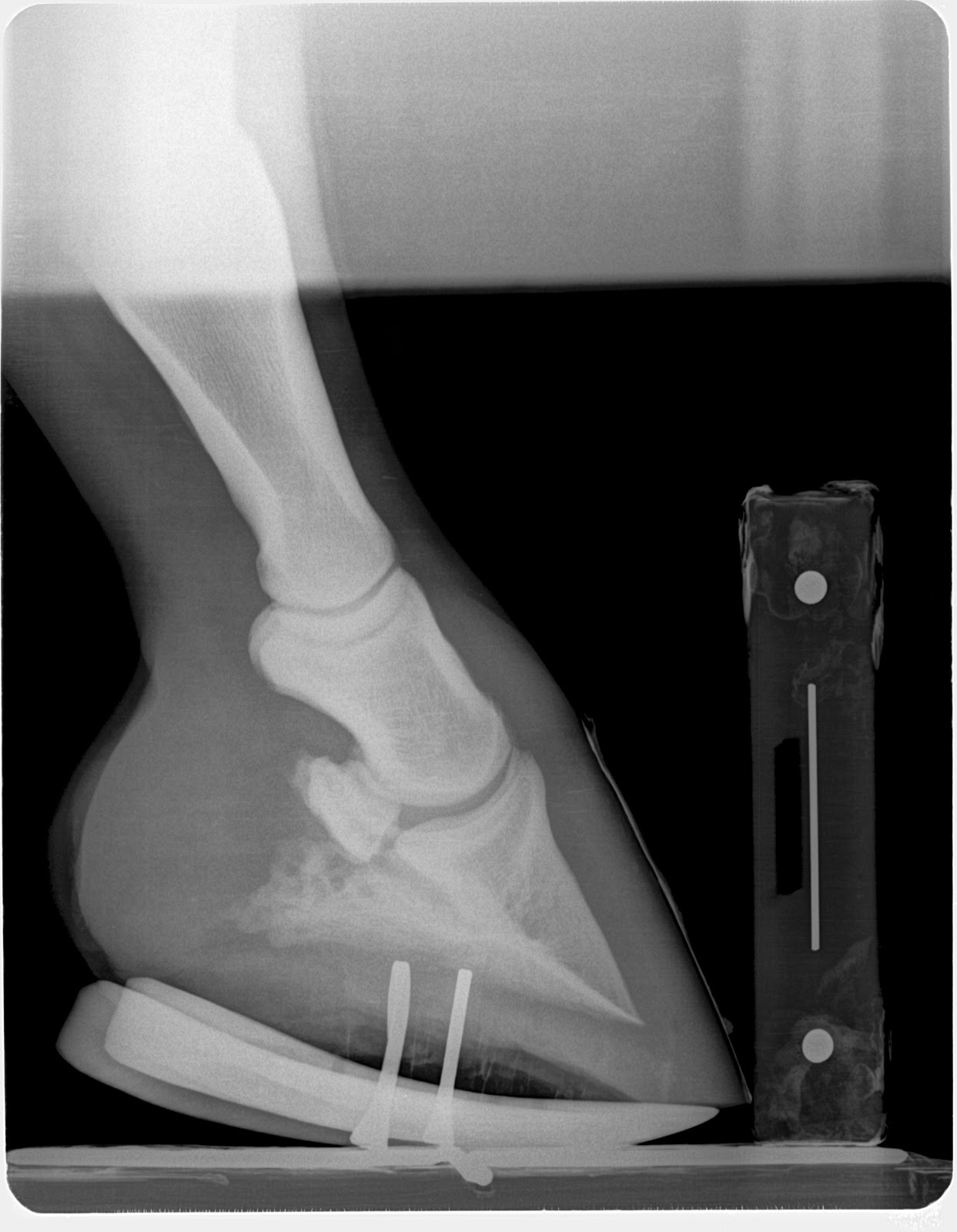
I do not recommend a tenotomy for every laminitis case and only do so if the venogram shows the circumflex artery at or above the level of the tip of the coffin bone as described by Dr. Ric Redden. However I do recommend considering the forces applied by the ddft to the coffin bone and often use "mechanics" (rockering/wedging) to lesson the tension on a failing system to aid in re-establishing vascular supply.
Below is several follow up images and short discussion of each case. Also look back at the previous blog entry for initial images and therapeutic shoe applied. October clinic images link
White line disease Case: Sole depth improved by 4mm but white line lesion failed to grow down at same rate and decision was made to remove hoof wall to expose oxygen and allow cleaning. Owner reports that he is running around like a youngster again and is more comfy than is has been in a long time.
 |
| 6wks post intial rocker rail note 4mm increase in sole depth in a horse that hasn't grown any sole in years. |
 |
| Reset image |
 |
| First image Oct 6 pre shoe |
 |
| Hoof wall resection to allow cleaning and oxygen to penetrate |


Club foot case: This horse lost the rocker rail shoe applie to the foot opposite the grade 3 club (which is also a club) and regular farrier applied a flat steel keg shoe to keep foot protected. Note the horn lamellar zone divergence. One could call this rotation which would be non specific. The divergence is created by the club syndrome stretching to lower horn to bone attachments. This is confirmed by evaluating the dermal-epidermal junction and measuring the horn zone compared to the lamellar zone. If the lamellar zone was larger than the horn zone one could conclude a laminitis as this is lamellar swelling. In this case it is chronic stretching of the lamellar bone secondary to the constant pull of the deep digital flexor unit.
The Grade 3 club grew more sole in the rocker rail than did the lower grade club in a flat shoe. This information tells us that placing the tendon sling in freedom with the rocker shoe allows better nutrient and blood circulation through unloading of the sole via reduced deep flexor tension. We placed the grade 2 club (Left Front) in a rockered trim with rockered steel keg shoe to also place the tendon sling in release. We will be to see a more rapid sole mass recovery in this hoof as well at the next reset. Owner reports excellent comfort and has adjusted very well to the new shoeing approach.
 |
| Pre shoe radiograph Oct 5 |
 |
| Left front shoe that regular farrier had replaced with flat keg shoe for protection |
 |
| Rockered keg shoe |
 |
| 6 wks post rocker rail application additional 4mm of sole and cup starting to form. All this due to unloading of the deep flexor pull |
Chronic Lamintis case: Farrier was a student and he reports horse is moving very nice. Horse was able to stand comfortably for each shoe reset. Turning and moving very nicely.
 |
| Pre Rocker shoe oct 6 |
 |
| 6 weeks post rocker rail with addition of 4mm of sole and less bulge of sole at apex of frog. |
 |
| Oct 6th pre rocker |
 |
| Left front 6wks post rocker rail. Rocker shoe was removed prior to getting a radiograph. Added 5mm of sole |
 |
| Nov 17th reset with rocker rail. |
 |
| Post nov 17th reset rocker rail. |
Navicular case: Owner reports she was able to work a pattern for the first time in 2 years. The Owners farrier was present and we helped him reset the rocker rails. We plan to maintian the higher palmar angle for the next shoe cycle then began to lower the mechanics/palmar angle. I expect to achieve similar comfort with lower mechanics as the horse remained comfortable even with losing a few degrees of palmar angle secondary to growth. The history is very important here. If horse became more lame at the end of the cycle as the palmar angle decreased, this tells us the hot spot becomes loaded at the lower palmar angle and may require a longer period of higher mechanics.
 |
| RF pre reset on nov17th |
 |
| Post shoe nov 17th |
 |
| Post shoe nov 17th |
 |
| Pre shoe reset on nov 17th |
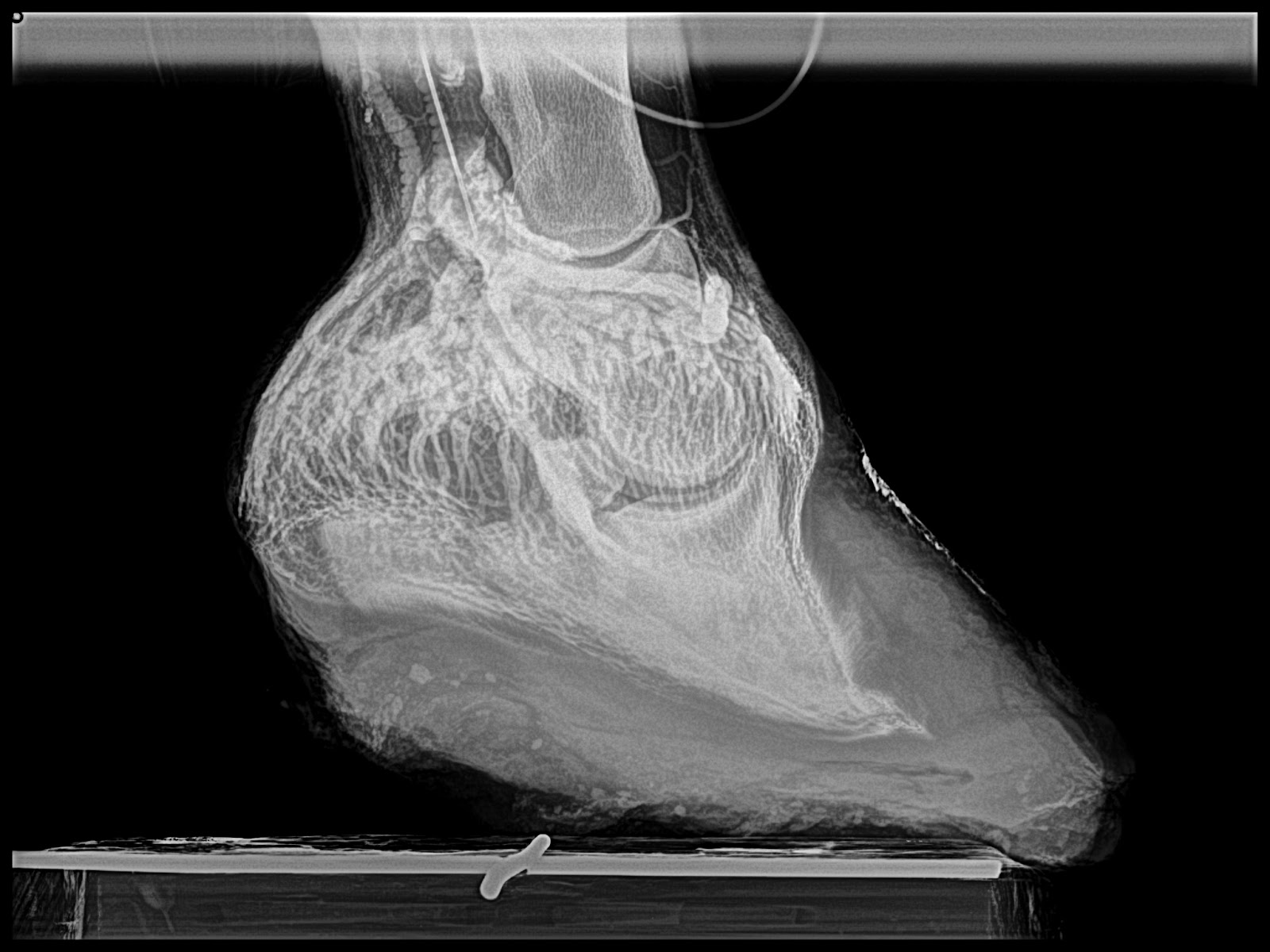
6 month chronic laminitis case: Owner reports horse is very comfortable, has a much better appetite and very willing to move freely. This case demonstrates the importance the deep digital flexor tendon force applied to a failed lamellar bone. With the loss of the lamellar suspension of the coffin bone, it is allowed to compress the sole at the apex of coffin. No blood, No growth and recurrent abscessation as has occurred in this case. The fragile rim of the coffin bone becomes loses its blood supply and acts like a foreign body. I haven't been able to achieve this level of success with any other approach. Doubling sole depth from 10mm to 20 mm in a matter of 6 wks in chronic laminitis is astonishing.
I do not recommend a tenotomy for every laminitis case and only do so if the venogram shows the circumflex artery at or above the level of the tip of the coffin bone as described by Dr. Ric Redden. However I do recommend considering the forces applied by the ddft to the coffin bone and often use "mechanics" (rockering/wedging) to lesson the tension on a failing system to aid in re-establishing vascular supply.
 |
| Immediately post derotation and deep flexor tenotomy oct 6 |
 |
| Note the rapid growth of sole at dorsal portion of hoof and loss of palmar angle. addition of 10mm of sole |
 |
| Post reset to re establish a zero palmar angle with the shoe. This is necessary to prevent over correction resulting in a negative palmar angle |
 |
| Immediately post derotation and deep flexor tenotomy on oct 6 |
 |
| 6wks post derotational shoeing and deep flexor tenotomy. No reset required as even sole growth is occuring and resetting the shoe does not add any benefits mechanically. |