Welcome, About us

Hello and welcome. My name is Sammy L. Pittman, DVM and I am a veterinarian, farrier, and horsemen with a great interest in the field of equine podiatry. My wife and I own and operate Innovative Equine Podiatry and Veterinary Services in Collinsville TX. My passion lies within the health and well being of the hoof to better serve your equine companion. With so much lameness attributed to the lower limb many horses require an out of the box approach to achieve the success desired.
Give us a call and we will be glad to help you in any way we can. Thanks so much.
I will be discussing different Cases and thoughts from our world with the horse. Feel free to contact us via text or call at 918.235.1529 or send an email to iepvs11@gmail.com. Thank you for reading and enjoy

Tuesday, July 26, 2011

Lobo the Laminitis case.


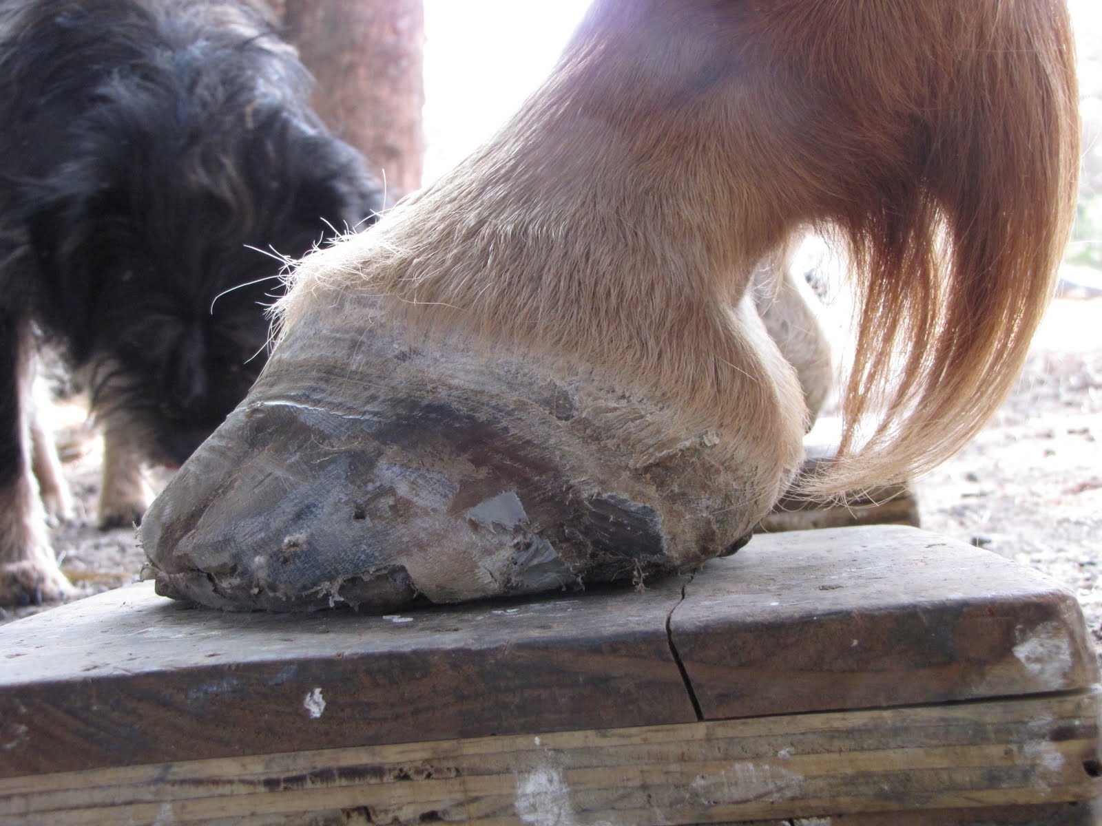
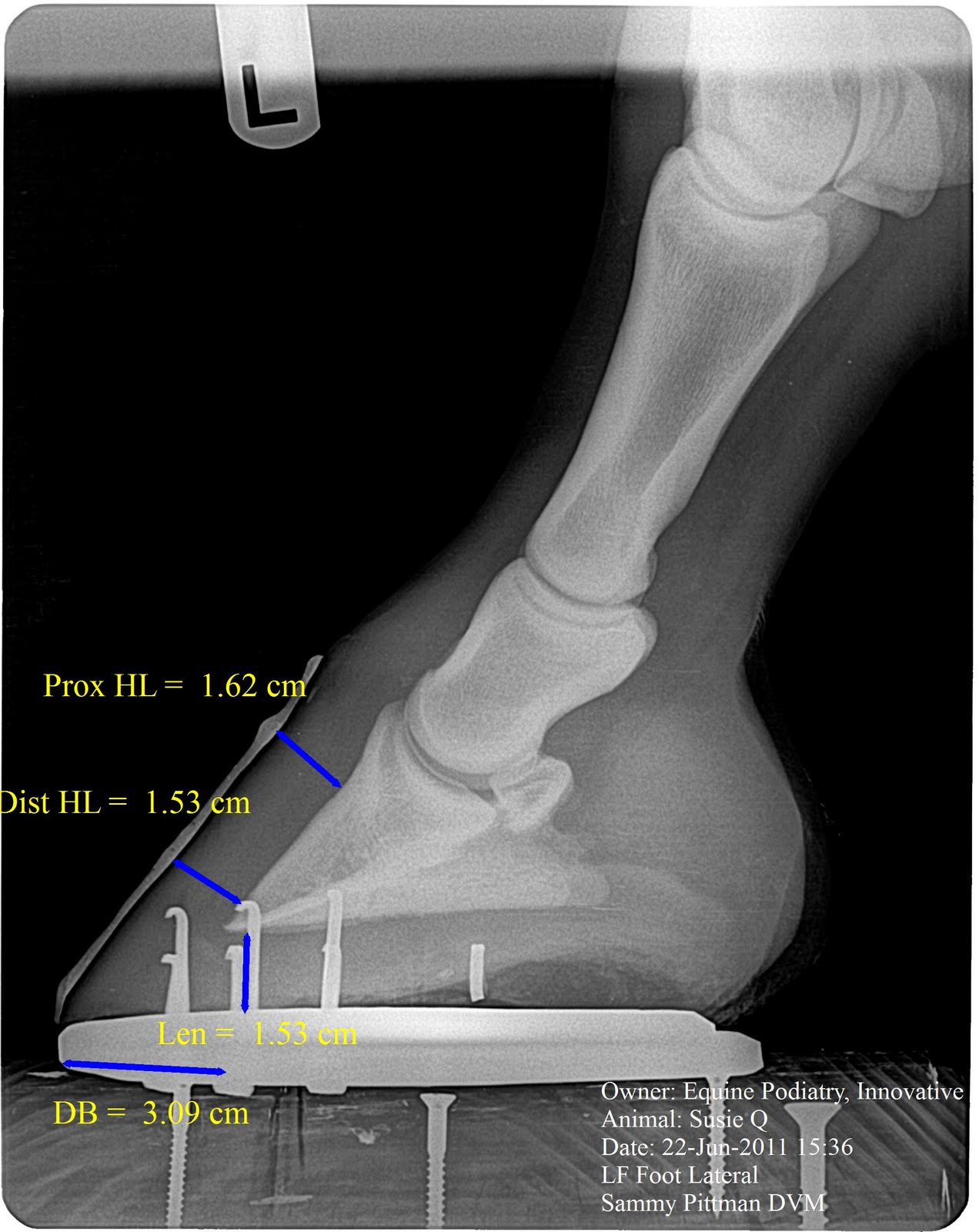
Welcome again,  This is a case of acute laminitis that we were called to look at late on the evening of May 29, 2011.  History of colic episodes the previous couple of days but now not wanting to stand and when does stand has typical laminitis stance.  Upon examination normal foot conformation no ridges but large bounding pulses noted in all four feet.  Placed in Nanric modified ultimates which raise the palmar angle 18-20 degrees and Lobo immediately began to show signs of comfort with licking of lips, less distress and standing up squarely on fronts.  He was still reluctant to move in a normal fashion.  Baseline radiographs are taken on this evening and have soft tissue parameters within normal range.  This is where many hoof care professionals are confused as no signs of bone displacement or rotation has occurred, but it is still very important to support this foot mechanincally with wedging to unload effects of the deep digital flexor tendon as the vascular compromise has likely began.  The idea that if there is no rotation it is no laminitis/founder does not hold true. This will also show how important radiographs on day 1 of  exam are so valuable when compared to the next visit 5 days later.  A baseline venogram was not performed as finances where limited at this point.


We returned to visit Lobo five days later and find he has been laying down the biggest part of the time which is probably to his advantage as all load is off feet and better circulation is allowed.  Radiographs on this day show significant soft tissue parameter changes with large increases in CE, H/L zones and decrease in sole depth.  Palpable ledge is noted on both fronts at the coronary band that is consistent with a sinker.  We performed a venogram at this point at no charge to client to further increase our knowledge of this case.  The changes in soft tissue parameters indicative of a moderate sinker are confirmed with the venogram with no perfusion at the coronary waterfall, face of coffin bone and sole under tip of p3.  Note even in this severe case the heels remain great perfusion.  The unloaded view is taken with the limb being held up which gives us an idea of what the perfusion may be while laying down with tendon and foot completely unloaded and some indication of what it would look like with  a tenotomy to completely release any action on the coffin bone. 

The venogram is performed in the modified ultimate which unloads the flexor tendon by 60 percent.  This also serves as a means to evaluate a certain therapeutic package.  If your plan is to restore healthy blood supply then you should be able to prove that the package is going to do that via the venogram.  This venogram suggest that the modified ultimate will not be enough to restore proper blood supply and that further mechanical release via deep digital flexor tenotomy would likely improve the situation as the sharp border of the coffin bone is cutting through the sensitive solar corium and its vital blood supply.  Several methods to prevent the bone from displacing are practiced but few have been confirmed and followed with serial venograms and radiographs to prove that they are unloading the circumflex artery.  The heart bar shoe is a positive force applied to the frog in order to antagonize the displacement of the coffin bone, but think about the tissue between the rigid heart bar and the coffin bone.  The solar corium with its blood supply that makes the horny sole that we can see and touch.  Obviously it has helped many cases but I think a further study as to it's affects on foot perfusion in a laminitis scenerio via serial venograms and radiographs is warranted.  Below is the radiographs and venograms from the second visit 5 days later.  I will also post a normal venogram for comparison.
NORMAL VENOGRAM FOR COMPARISON TO LOBO'S


We were unable to perform suggested derotation and tenotomy as financial constraints did exist.  Owner opted to give Lobo more time as he was laying down which is protecting the vital blood supply.  Contact was  made with owner approximately 6 weeks after second visit and Lobo was not improving and he was ready to euthanize and I offered to take Lobo to further his treatment and if not able to be successful I would humanely euthanize if needed.  Lobo was transported to IEPVS and new radiographs and venograms performed.  Notable hoof wall growth had occured and was surprisingly very close to even from toe to heel.  Moderate improvement in venograms in areas of the coronary band, face of p3 and tip of p3.  Considerable displacement of the circumflex still exist and very severe compression of vascular bed under apex of p3 is still present.  Note the changes in the soft tissue parameters on the lateral films.  Note the lucent zone that indicates the level of separation of the horn/lamellar zone.  Despite improvement of venogram, more mechanical release in the area of the circumflex  and solar vascular bed is need and derotational shoeing and tenotomy was performed.  Note the red line is the trim line and guide for shoe placement.  The shoe is a 5 degree rail shoe with a trailer welded in to prevent the toe from tipping up after DDF tenotomy.  The shoe is placed at zero palmar angle with a minimum of 20 mm space below p3 and the 5 degree helps prevent painful subluxation of the coffin joint.  This shoe is atraumatically applied with adhesive and nylon strips.  Two part silicon rubber is used to prop shoe up and apply caudal solar weight distribution.


POST TENOTOMY RADIOGRAPH

TENOTOMY RAIL SHOE, POST TENOTOMY RADIOGRAPH

Lobo has went from laying down 90 percent of the time to standing 90 percent of the time.  Attitude and appetite have greatly improved.  The plan is to recheck venogram and lateral radiographs in 2-3 weeks after tenotomy.  I will gladly post those so we can all learn from this case.  With the amount of bone remodeling that has already occurred to the fragile thin rim of coffin bone it is less likely we will have as good of a response as we would have if derotational shoeing and tenotomy where performed in the important window of opportunity at the second visit.  However, I do expect a great improvement in the hoof growth and patient comfort.  The amount of bone resorption will be what prevents Lobo from returning to a previous level of perfomance.  We will have to wait and see.

I would like to thank my Lovely wife for helping and enduring the  many crippled horses I drag to home to try to fix and learn from.  It is vital to my education to have cases like this. I would also like to thank Brendan Frost for donating a Sunday afternoon to help me shoe Lobo.

Stay tuned for more exciting podiatry cases!!!!!!!!!!!!!!!!!!!!!!!!!!!!!!!!!!!!!!!!!!!!!!!!!!!!

Sunday, July 24, 2011

Follow up on the Severe White line Disease case 7/22/2011

Hope everyone is staying safe in this record warm weather here in the US.  For those of you not suffering from the heat, your lucky.  Well the horses don't care how warm it is they still need there hoof care!  I revisited the white line disease case this past Friday.  We have continued accelerated hoof wall growth without cracks.  The Right front which is the more upright still has signs of fungal invasion despite mechanical unloading noted by the lucent zone in the Horn component of the H/L zone.  This would likely suggest the pathogen is invading new growth.  Left front shows continued improvement in all aspects with good hoof wall growth and sole depth recovery.  Sole growth has been slow to recover but is measurably increasing at this point.  I feel that higher scale mechanics (ie more rocker or deep digital tendon release) will be required for continued success in the right front due to this being the more upright foot and is under more deep digital flexor muscle pull.  At the last visit I instructed the owner to place on a weight control program with only enough alfalfa pellets to get the 100mg biotin and vitamin and minerals in.  Significant weight loss has occurred and will help the overall success of this case as the obesity could increase insulin and decrease the amount of circulation to the lower limbs further decreasing hoof quality and quality.  A great improvement is noted in hoof structure with loss of flares and a tighter new growth coming down.
    I elected to remove more dorsal hoof wall in area's that cavities existed and pack with a mixture of pine tar and oakum versus cleaning and packing with white lighting gel.  Below are updated photos and radiographs.  Read the captions for further information regarding individual images.
Increased sole depth but lesions from fungal invasion have remained

Very first radiographs
Good improvement in sole depth and new growth without fungal invasion


right front with 1/3 of new hoof growth.

Left front with almost half of new hoof growth without cracks.


Shoes are attached with a few nails into hoof wall then 3-4 next to hoof wall and superfast adhesive is used to glue  nails to hoof wall.  A band of superfast is added across the front to attach the two sides.  This has been one of my tougher cases and I appreciate the opportunity to work on this difficult case and the commitment the Owner has made to her equine companion.  We still have several months to go but I feel we have made significant improvement.


    We have two cases in the barn right now that we have been working with and plan to post them here on the blog as soon as time will allow to put together all the images, time lines and thought processes.  One is a fractured second phalanx (short pastern) named Lila that is recovering nicely and the second is a newly acquired laminitis case that was acutely laminitic about 6 weeks ago.  We will be posting those soon so keep checking back.  I am also excited about attending Dr. Redden's In depth podiatry 201 course August 8-12 in Versailles, Ky with farrier and friend Brendan Frost.

CHECK OUT WWW.HEARTLANDHORSE.COM FOR THE ONLINE VERSION OF THE HEARTLAND HORSE TRADER FOR MY FIRST ARTICLE EVER PUBLISHED!!  Look for it in all your local feed stores, tack shops and shows.

Stay cool, but most importantly Stay Fresh,,,,in your knowledge.

Tuesday, June 28, 2011

Backyard mare project update with venograms

Well it has been a while since we looked in on our backyard mare project.  We found some time to radiograph and update her venogram.  She has grown to a little over 17mm of sole depth and a notable return in solar papillae.  She has just been in the rockered 4pt shoe and maintained on pasture and trailriding.  I have placed her back in a more traditional flat steel shoe and placed in a fashion that would commonly be applied.  I theorize that this breakover is too long even on the day I placed it and will be growing ever longer throughout the shoe cycle.  The flat shoe will once again solidly engage the deep digital flexor tendon in which will begin to compress the solar corium and decrease sole depth over the next 2-3 months.  We will continue to radiograph and follow to see what happens.  
Very first venogram 3 1/2 months ago, for comparision


Note the return of the solar papillae with added foot mass.  This is the reason that Dr. Redden  claims 15mm is an adequate sole depth, as is requires 15mm to have a normal healthy venogram with good vascular depth and these papillae.  There is a peculiar absence of contrast that is there from an unknown cause, maybe an abscess brewing creating internal pressure, pushing the contrast out????
Very first raidograph, note the very thin soles.  

Here is a shot just before the removal of the shoe and venogram.  Note the massive increase in sole depth since we began.

Here is a shot after venogram and I placed her in a more common flat steel shoe in a manner in which I would have placed prior to a changing of my mind of how forces interact in the foot.  The DB (digital breakover) is 30 mm. This will continue to lengthen over time and reaching a likely 40-45mm by the next shoeing cycle, possibly.  I also surmise that a gradual reduction in sole depth will also occur due to the long and every increasing DB and increasing forces from ddf on solar corium and dorsal hoof    decreasing the circulation.
solar shot of flat steel shoe.

lateral view of flat steel shoe.  For those of you who know me, know that this was very difficult for me to do.  HEHEHEHEHEHe
Keep checking back for further updates on all our cases and a new one still to come is project Lilla.  She is recovering nicely from a fractured short pastern bone.  I will post here case study as soon as time will allow.  Please feel free to contact us here in Tulsa, Ok via text/call at 918.235.1529 or email at innovativeequinepodiatry@hotmail.com.

We are getting prepared for several upcoming events such as:  July 9th a reduced cost coggins clinic at animal health supply in Tulsa Ok, Aug 8-12th Dr. Redden's 201 podiatry course at International Equine podiatry center in Versailles Ky, 1-2 local demonstrations of radiological and external exam of the hoof and soft tissue parameter measurements, and of course the In depth podiatry lecture and demo with Dr. Redden here in Tulsa October 14th and 15th.

We thank you all for your support.

Friday, June 17, 2011

White line disease case and upcoming reduced price coggins clinic.

We have been quite busy and I just haven't had the time to catch up on the blogging. We a planning a reduced price coggins clinic at Animal Health Supply on July 9th from 9-12.  If you have any questions please call 918.235.1529 or shoot us an email at innovativeequinepodiatry@hotmail.com.  Cost of coggins test will be 15 dollars and you will also receive a 10 percent off coupon for Animal health supply for that day, good for anything but dog, cat and horse food.  Hope to see you there.

I am also posting some Follow up radiographs and pictures of the White line disease case we have been following.  This case is still progressing but not as much progression as I would like to see on the right front.  We have good hoof wall growth but not as much sole depth as I would like to see.  With the onset of good green pasture and the potential for some insulin resistance in this case could have played a role in slowing of hoof growth.  We instructed to reduce the amount of pasture time and absolutely no grain products.  Both hooves are becoming much tighter and healthier.  I removed more dorsal hoof wall to allow cleaning and treatment.  The left shows significant new hoof growth without fungal invasion. noted by the measurement on the radiograph.  We reset the Rocker Rail shoe with frog plate.


Note new hoof wall growth that is nice and tight without a crack.

New growth without a crack


I am happy with the amount and quality of hoof wall growth.  I would like to see a faster sole depth recovery but it will come.  This didn't happen overnight and we probably will not fix it overnight.

Keep in mind the Upcoming In Depth Podiatry Clinic with Dr. Ric Redden October 14-15.  I have some new cases that I will likely be posting on.  Keep on the lookout for Project Lila a barrell/pole horse with a fracture second phalanx (short pastern bone). She is doing well and will likely make a good recovery.  I will post images and discussion as soon as there is more time.

Tuesday, May 24, 2011

Just some thoughts for today.

Hello everyone,  we are pleased to announce we have been granted a distributorship for Nanric.com.  This will allow us to sell Nanric products to you at a comparable price as their online pricing but save you money on shipping cost.  Please call us or email us at innovativeequinepodiatry@hotmail.com if you have any questions or would like to try some shoes.  


We had decent turnout for our first coggins clinic last saturday and we are planning a couple more in the next 6-8 weeks.  I am also planning 2-3 half day demonstrations regarding radiographic and external evaluation of the equine hoof.  These will be low cost or potentially free presentations open to veterinarians, farriers, trainers and horse owners.  I am trying to give a little taste of what you will be exposed to with Dr. Redden's in depth podiatry lecture October 14-15 here in Tulsa.  Please be on the lookout for those dates and places, and if you are interested in hosting demonstration please let me know.  


We made a few changes to the Blogsite to include an up to the minute news reel from the thehorse.com and some videos we have on youtube.  Keep checking back for the latest equine news especially regarding the lastest information on EHV outbreak.  If you have any questions regarding the health and wellbeing of your equine family please do not hesitate to call.  


We are rechecking the severe white line disease case this weekend to be sure to look for a new blog regarding updated cases next week!


God Bless and keep all the tornado victims in your prayers.

Wednesday, May 4, 2011

Upcoming Podiatry Clinic with Dr. Ric Redden.


Go To Our Website, http://www.innovativeequinepodiatry.com/ to download a brochure to fill out or click on the link to register online.  See you There. 


We have many of the details completed and it is a go for hosting internationally renowned veterinarian/farrier Dr. Ric Redden for a very intense lecture and demo.  You will be exposed to a very detailed evaluation of the equine hoof and its mechanical forces.  Don't miss out!

Please Call, text or email if you have any questions.   918.235.1529, innovativeequinepodiatry@hotmail.com
We are getting prepared for the Magnolia Tx clinic and I should have some good photos and images from this weekend.  Hope to see many of you there.

Monday, April 18, 2011

Follow up on the Severe White line Disease case

Hello readers, I hope this finds you well.  It has been a very dry Spring here in Oklahoma and fires are really causing troubles in many areas.  We definitely keep the firefighters in our prayers.  We had a busy weekend in the podiatry world.

On to the Case.  A very good response is noted since our last visit.  I consider a good response in a severe white line disease to be new hoof growth without fungal invasion.  The hoof defect noted in the soft tissue lateral radiograph is growing down.  The left front has 13mm  of new hoof growth with out cracks.  There is still  uneven growth rings from toe to heel but greatly improved.  Sole depth has increased but is still considered severely thin soled.  If you look at the upper portion of the barium paste you can see the new hoof wall growth that is growing down more parallel to the front/face of coffin bone/P3.  Patient has been in a small paddock and fed Biotin 100 from nanric.com daily.  I will be posting photos and radiographs from first visit and this recent visit.
first day pre shoe


first day post shoe
Before glueing 5weeks after initial visit.  Added a little more mechanics/belly/rocker to shoe.


With Superfast,  Due to poor quality walls I like to add a little extra holding power.  I drove nails but left long and create loop in end for glue to attach to
Initial visit post shoeing radiograph

Recent visit 5 weeks later, Notice the hoof wall defect growing down without fungal invasion and more parallel to front of coffin bone.  I also added a little more rocker to shoe to increase mechanics that allow further unloading of deep digital flexor tendon.  This image is after reset of shoe.
Left front day one

reset Note new growth without cracks in the upper hoof wall 
Now on the Right Front.....................................................................................................................................................
Day one
day one right front
Post Shoe day one with superfast.

Post shoe at 5wk reset.

5 week reset pre superfast.

After super fast

Day one post shoe radiograph

5 week post shoe radiograph.  Notice new growth at upper coronary band that is growing more parallel with front of coffin bone/p3

 So to restate the purpose of the use of the Rockered Rail shoe.  The Rails give us 5-7 degrees of heel elevation and the added rockering adds a self adjusting air wedge to allow further unloading of the tension in the Deep digital flexor and decreasing the pull at the inner layers of the horn wall.  The rocker action is also thought to increase the circulation of the entire foot which will enhance hoof growth and quality.  With the rocker action Digital breakover is greatly reduced.  I also added in a positive pressure frog bar to help prevent buttress/frog prolapse and spread weight distribution across the back of the hoof.

The most important aspect of why the rocker shoe is successful is it's ability to greatly reduce the action of the deep digital flexor tendon.  The deep digital tendon and it's action is the "big dog" force in the Hoof.  You must consider it's action and the forces it applys to the coffin bone and the space around it.    It pulls tension on the bone to hoof connection and a downward compression force on the solar corium below the tip of the coffin bone.  We must always first consider the action of DDF  and all other aspects of shoeing will be secondary to that.  I like this approach versus wedging alone as it can be difficult to acheive this much DDF tension release and breakover reduction with wedges.  Additionally the rocker motion greatly enhances the healing environment and speeds up recovery.  The addition of the superfast  at the quarters and over the toe adds stability  and connection of the whole hoof once again.  I do not feel it is required but does help hold the shoe longer.

Just Got off the phone with Dr. Ric Redden and things are coming together for the October lecture and demonstration.  I will have brochures ready within the next week.  October 14 will be an all day lecture and then on the 15th will be a demonstration at the 181 ranch in Bixby Oklahoma.  It will be a very informative seminar and hope you can make it.

Thanks for reading,                    Sammy L. Pittman DVM

Wednesday, April 13, 2011

Follow up Venograms and Radiographs of the Backyard Mare project.

. Hello everyone, I hope you are enjoying this site and potentially learning something as well.  Please feel free to ask any questions in the comment segment.  So we are just a little over four weeks into this shoe cycle with the low score mechanical rocker shoe.  In the plain film soft tissue farrier friendly radiograph we can measure right at 14 mm of sole depth in the left.  We started with 8mm of sole depth when we pulled her out of the pasture with bare feet and slightly tender on the rocks.  She has been ridden fairly regular which does slow down foot growth.  Again, what I am attempting to demonstrate is how the rocker shoe application enhances the  foot mass noted by increase in sole depth and the accompanying venogram changes. In my experience and the experience of many others, the rocker shoe is the fastest method available to regain foot mass and quality.  We are getting close to normal sole depth  of 15mm after 4wks in the rocker shoe.  I find that many foot ailments and lameness' improve drastically with added foot mass.
 The first image is today's and the second is the initial radiograph March 8, 2011.  Notice the great improvement in sole depth.
The Most notable change here is the terminal papillae are getting longer.  These are the little finger like projections.


In this view the terminal papillae continue to become longer and  more robust.  This will continue as sole depth increases.  When we achieve a healthy sole depth of closer to 20 mm we should have a very prominent terminal papillae and a vascular depth of around 10 mm.  


Below I am going to post a recent venogram on a chronic laminitis case.  This gives something to compare to.  Note the severe lack of contrast over to face of the coffin bone and below the apex of the coffin bone.  Just to refresh the memory the white tree branch like things you are seeing are the arteries and veins within the hoof.  In the image below there is no blood flow below the coronary band in the front (to the left).  There is still slight filling of the terminal arch which is supplying the coffin bone its nutrients and is keeping it from completely dying.  There is considerable bone resorption as almost 1/2 of the coffin bone is gone due to the lack of normal blood supply.  
 Keep checking the blog as next week I will be rechecking the Severe white line disease case that I have already posted.  We will be repeating radiographs and evaluating response to the rocker rail application. 

We hope everyone is enjoying the Spring.  

We are in the planning stages of hosting Dr. Ric Redden of International Equine Podiatry Center of Versailles, Ky for a 2 day lecture and live demonstration on October 14 and 15.  I will post the details when we get them nailed down.  

We will also be traveling to Magnolia, Tx to help renowned farrier Jimbo Stewart put on a clinic demonstrating radiographic technique, how to treat common foot problems and to read radiographs.