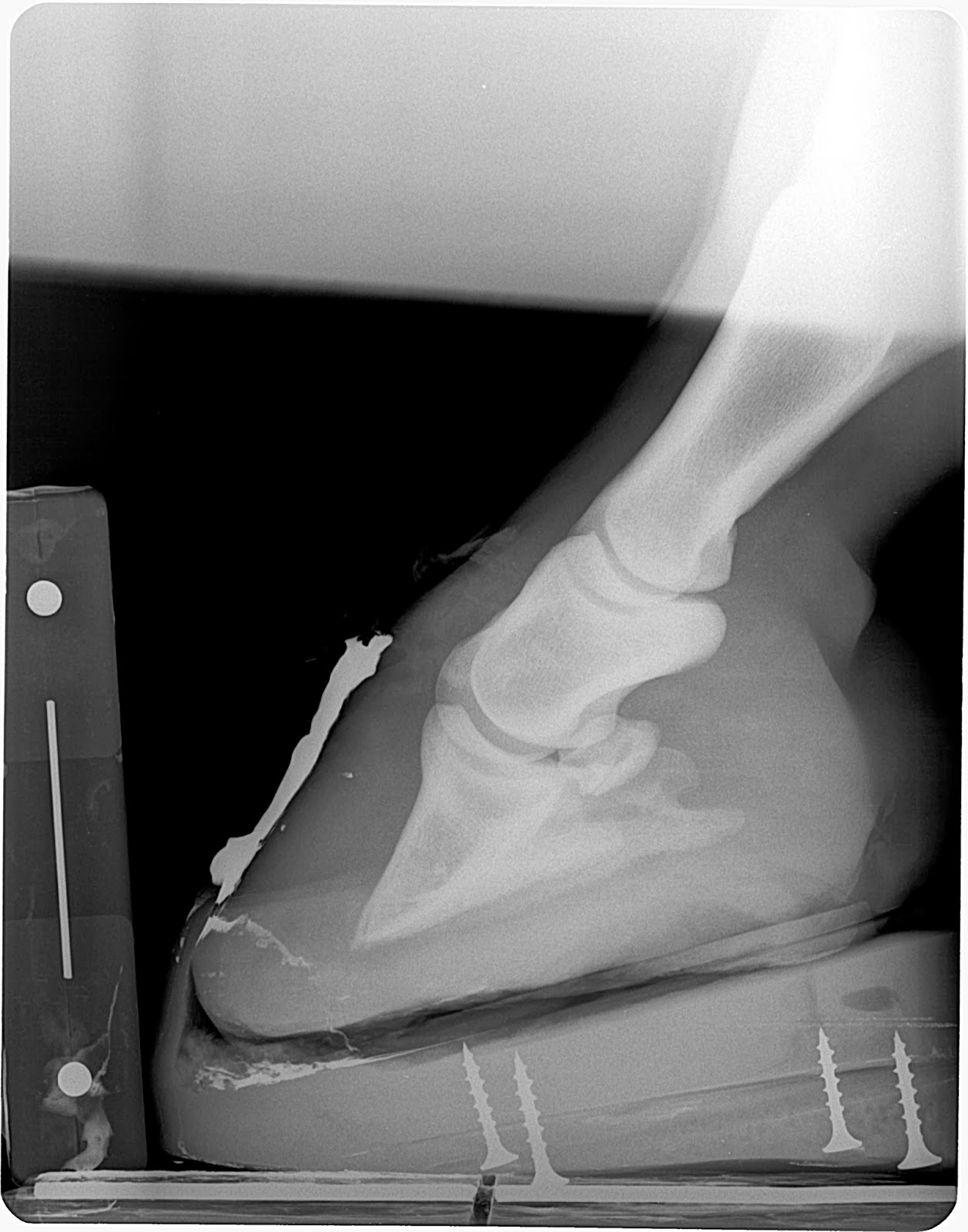
Welcome to 2014! I wanted to review some of my laminitis cases that have proven very successful with regards to quickly adding sole mass and demonstrating an even hoof wall growth from toe to heel. A couple of cases will also demonstrate how quickly the venogram can change. Improvement in the blood supply is what we are all after.When you can demonstrate a quickly improving venogram study plus the quick addition of sole depth you can be a more positive about the overall situation. Success to me is rapid foot recovery and ideally reversing the damaging effects of vascular compression before it creates irreversible bone and soft tissue damage. Monitoring with venograms will show the level of vascular damage present and allows a quicker more accurate mechanical therapy.
For a review on soft tissue parameters measured on a podiatry style radiograph click here.
For a reference on a healthy venogram click here.
The first case is Rocky. Rocky was first examined about 3 months after the initial insult and was well past the ideal time to completely avert any bone change. Note the big divot out of the tip of the coffin bone caused by a severely displaced circumflex artery and terminal papillae is supplies. This chronic history, severe coffin bone displacement and venogram indicated the need for a deep flexor tenotomy (cutting of the tendon) after derotational shoeing.
Note the quick addition of sole mass and a decrease in the amount of rotation within the hoof capsule. Loss of rotation is not the goal but a common finding after changing the load dynamics by cutting the tendon. This places a majority of the load towards the back of the coffin bone and can push the tip up in many cases to reduce the amount of rotational displacement and unload the circumflex under the rotated tip of the coffin bone. Left column is the day of surgery and the right column is 30 days later. Note the rapid addition of sole under the tip of the coffin bone. The 3 months prior the hoof wall growth was greater in the heel than in the toe area which is very common with laminitis. This is secondary to the vascular compression in the front half of the foot. This is confirmed in the above venogram. The blood supply to the coronary band should be much fuller than demonstrated here. After the tenotomy the hoof wall began to grow more even as we have unloaded the forces applied by the tendon and allowing a reperfusion of blood to these vital areas.
The images in the left are post shoeing radiograph from the 30 day post tenotomy visit and the images on the right are 30 days after that or 60 days post tenotomy. The inital shoes are glued on and are usually left on for the first 10-12 weeks and many cases are barefoot at that time. This case was growing so rapidly and to properly manage the palmar angle (prevent from getting into the negative zone) the shoe was removed, the foot trimmed and very lightly nailed back on parallel the the wings of the coffin bone. Again note the amount of sole depth recovery within this 30 day period.
The bottom two images are 90 days post tentomy. This horse is comfortable barefoot and can maintain a zero to slightly positive palmar angle.
Plan is for this horse to start hand walking daily for 5-10 minutes just to get him out of the stall. Recheck at 4-6 week intervals with radiographs to insure continued foot mass recovery and maintenance of the palmar angle. This horse may very well be able to do some light riding in another 6-8 months with some turnout. Because of the severe bone remodeling that had already occurred I am hesitant to say he will return to 100 percent of what he was prior lamintis but can have a good quality of life.


Case #2 Gracie
Gracie had been guilty of getting into the owners bird seed and dog food and was a touch overweight. Surprisingly fairly sound and would only rock back on hind quarters in turn. A considerable amount of coffin bone displacement had already occurred which indicates the syndrome has been rolling for some time. Owners reported some pain over the last 4 weeks only. Note the distal divergent horn lamellar zones and loss of sole depth.
Placed in Nanric modified ultimates the performed a venogram. Venograms in the left column are the first exams and the right column are venograms performed 9 days later. Note the improvement in the vascular structure around the tip of the coffin bone. This is secondary to the wedges unloading the tendon tension by decreasing the distance from its origin to insertion with the coffin bone. This allows the load to be transferred to the heels an back of coffin bone. I also measured a 3mm increase in sole depth in this short period. This is likely due to the unloading of the solar corium directly under the tip of the coffin bone. Think of placing a clothes pin on your finger smashing is flatter. It will measure a greater thickness once the clothes pin compression is removed and the tissue is once again filled with blood.


Below are images that are taken 30 days after placement in the nanric modified ultimates placing the palmar angle at approximately 20 degrees
Below are images that are 90 days in the ultimates wedges
This demonstrated a rapid change in the vascular pattern with the addition of the wedging and did not require a higher level of deep flexor tendon relief as the previous case in which the tendon was cut. Just placing a little slack in the tendon system is often all that is required to unload the vascular supply in important compromised areas in the front of the foot and directly under the tip of the coffin bone. This horse will then be transitioned to a rockered 4pt rail shoe that will continue to offer a greatly reduced load on the flexor tendon, solar corium and the lamellar attachments. The level of rocker/mechanics will be slowly lowered as long as continued soft tissue response is noted.
Case #3 is a mustang that suffered lamintis in all four feet. The fronts required a deep flexor tenotomy as the circumflex artery was displaced above the tip of the coffin bone and no contrast is noted below the tip of the coffin bone. The hind venogram was also compromised but to a lesser degree. The fronts where shod with a derotational tenotomy shoe followed by a deep flexor tenotomy and the hinds were placed in the ultimate wedges. A follow up venogram performed on a hind foot demonstrated a positive improvement and suggest that a tenotomy is not needed at this time and supports continued use of the wedges. One can already see the addition of sole depth in this short 10 day period in the fronts and hinds.
On the left below is a venogram of the left hind on initial exam and a follow up venogram 10 days later while wearing the modified ultimates placing the palmar angle at 20 degrees. Note the significant improvement in vessel filling over the coronary band, the more normal appearance in the circumflex junction and return of solar and terminal papillae.
Below in the left column is 10 days post wedging and 30 days post in the right column.
Below are the hinds 60 days post placement into the modified ultimates.

These are just three cases from this summer and fall that demonstrated the quick response I am looking for. Understanding the mechanics relative to the deep flexor and its role in a failing lamellar bond has proven very beneficial in my practice. Monitoring the failing system with serial venograms will inform you quicker than plain radiographs. Instead of waiting around for 4-6 weeks to evaluate the response in sole depth and hoof wall growth the venogram will demonstrate the level of compromise days to weeks before any changes can be noted otherwise. This allows quicker changes in mechanical therapy and less irreversible damage. This prevents chronic pain and abscesses.
What do you consider a success? Patient comfort? What level of sole depth recovery in a period of time do you expect with your laminitis therapy?
Wishing you happy and prosperous new year.
All the best
Sammy L. Pittman, DVM


Thanks for sharing, I always look forward to your blogs. I am quite impressed by how helpful the venograms are in making decisions more quickly that will be in the horse's favor.
ReplyDelete