Welcome, About us

Hello and welcome. My name is Sammy L. Pittman, DVM and I am a veterinarian, farrier, and horsemen with a great interest in the field of equine podiatry. My wife and I own and operate Innovative Equine Podiatry and Veterinary Services in Collinsville TX. My passion lies within the health and well being of the hoof to better serve your equine companion. With so much lameness attributed to the lower limb many horses require an out of the box approach to achieve the success desired.
Give us a call and we will be glad to help you in any way we can. Thanks so much.
I will be discussing different Cases and thoughts from our world with the horse. Feel free to contact us via text or call at 903-718-0056 or send an email to iepvs11@gmail.com. Thank you for reading and enjoy

Monday, January 23, 2012

NANRIC Blog: Slideshow - Rockering a Rail Shoe

NANRIC Blog: Slideshow - Rockering a Rail Shoe: This slideshow of photos from Dr. Redden illustrates how to rocker a rail shoe.


Great slideshow of Dr. Redden Rockering a 4pt rail shoe which is used to treat many common fool ailments such as: chronic laminitis, navicular syndrome and thin soles.

Have a great week.

Wednesday, January 11, 2012

Update On Lobo the Laminitis Case.

 Previous Post on Lobo.  Click on the link to the left for the first post.  Lobo is responding very well to the Tenotomy performed back in July.  He continues to grow hoof wall at an even rate toe to heel and continues to add good dense sole.  I would not consider success if those are not occurring.  In evaluation of your laminitis treatment if you are still growing more heel than toe then you are not effectively addressing the mechanical problem. ie more tendon release is needed either with an increase in Palmar angle or likely a tenotomy.

I placed Lobo in Rocker rails as he began to have upright pasterns indicating the need for more mechanics (increase in PA), and due to the affects of the tenotomy which removes most of the load on the dorsal lamellar attachments and solar corium at the apex of the coffin bone but increases load through heels.  The only load is distributed through the bone column through the palmar (back) aspect of the hoof which over time will add some heel crush.  This is best addressed with the rocker or the buttress trim.

I (and Lobo) liked the increase in palmar angle but I was not fond of how he could rock back in soft footing.  This being due to the lengthening of the deep digital tendon post tenotomy.  I moved him into an aluminum buttress with a five degree rail, which accomplished my goal of a higher palmar angle and ability to remove all crushed heel and load to widest part of frog.  The buttress shoe trim involves creating a positive palmar angle by trimming from the toe back with the rasp tipped at a 5-8 degrees.  This is continued approxiametly the wings of the coffin bone ideally.  The heels are then taken at an angle parallel to the palmar angle to the widest part of the frog.  The  buttress wedge bar is made of 1/2 by 1 inch aluminum bent in to a U - shape to fit the width of the heels.  Forge a wedge shape to fit your trim. Grinding or hammering a recess in the bar for the frog will necessary in most cases.    I really like this approach for my post tenotomy cases when added mechanics are needed to maintain good digital alignment.  This will occur in more severe cases when vascular and bone damage occur secondary to laminitis insult that was either not appropriately addressed  in a timely fashion or so severe that any mechanics would not have changed things sucha as in severe sinker cases.  Lobo did not receive the needed tenotomy until 6 wks post laminitis and the initial insult was severe creating compromise at the circumflex. This is noted in the venogram obviously but also in the shape of the tip of the coffin bone.  You can see flipped up circumflex starting to remodel around the apex.
 Maintained in tenotomy rail then a four point trim.  This is when we noted a need for more mechanics noted by upright pasterns.
 Initially placed in Rocker Rail to increase PA and removed crushed heels.


Note the still compromised solar vascular supply with lack of papillae but nice remodelling of the circumflex artery.  


 This is the Left series which shows similar improvement as the right.    Good remodelling around the circumflex b ut poor solar and terminal papillae.  I feel that even with even toe to heel growth and good sole depth that unless solar papillae return with more healing and regeneration time then any chance of a performace career will be limited.

Points to remember:  Evaluation of the vascular supply is paramount to determine success of the device you are applying.  Success should not only be measured by comfort of the patient but I cannot claim success without even toe to heel growth and addition of 7-10mm of sole depth every 30 days.  If this is not happening one must rethink their approach.  Note we did not have a natural increase in Palmar angle we created is due to upright pasterns.  The upright pasterns can be secondary to continued low grade bone pain or even some heel pain.  Achieving better digital alignment allows load be directed through the middle of the foot in the direction that the first phalanx is pointing.

Lobo is now getting 4-6 hours of paddock turnout after five months of stall confinement with the first three in firm wraps over tenotomy site to prevent excessive scarring and a tenotomy rail shoe to prevent toe from flipping up or undue strain on tenotomy site in soft stall bedding.

Please comment, email (iepvs11@gmail.com) or call (9182351529) if you have any questions.

Thursday, December 29, 2011

Images from the October Podiatry clinic with Dr. Ric Redden, DVM

I hope all are having a great holiday season.  Kellee and I wish all of you the best in the new year.  We are looking forward to new podiatry cases and meeting new clients.

I am finally getting some time to post some images from the Redden clinic we hosted here in Tulsa Ok back in October.  We had a great time and had some good cases to work on.  Two cases I have continued to follow with radiographs and one with venograms.  Dolly the mild laminitis case we used as a venogram demonstration case I plan to post separately as a single case report after our next shoeing and venogram next month.  She was an interesting case and I have lots of images.  She is doing well and growing nice foot mass.

I can't thank Dr. Redden enough for spending his time with us in Tulsa.  Anyone that may be interested in attending future clinics in Tulsa please email us a iepvs11@gmail.com.

I will also be posting an update to Lobo the laminitis case with recent radiographs, shoeing approach and venograms.

Please look back in the archives to view many other cases and images.  I will be posting two more laminitis cases soon as well.

First case is a chronic navicular with rotational and varus limb deformities.  Goal is to increase tendon surface angle, Palmar angle and reduce digital breakover.


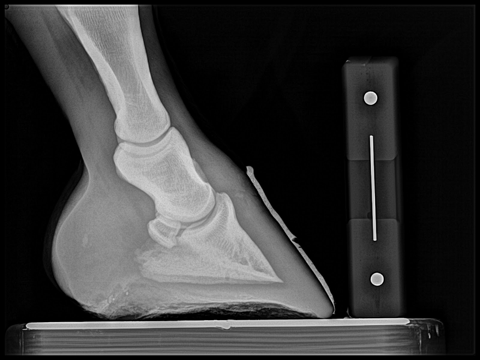
Below:  Quarter race horse that goes off after 150 yards or so and quits running.  4yrs old and has moderate navicular changes already.  Placed in Rockered Race shoe from NANRIC to increase TSA and PA. This will unload DDF/nav bone engagement and small osteophyte at dorsal aspect of coffing joint.


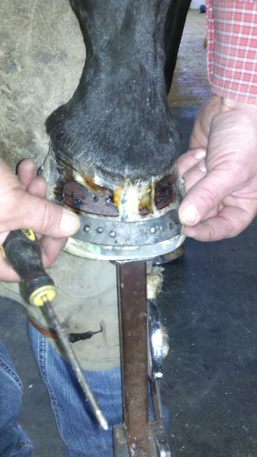
Next Case:  Older teenage trail and lesson horse.  He has been plagued with Navicular disease and is 2/5 lame on left front.  Immediate improvement is noted and absolutely no head bobbing at next reset.  The first three images are from the day of the clinic and the last two are pre shoeing radiographs from the reset.  I misplaced the day of the clinic rads for this case. 

This is is post trim with application of barium paste to accentuate the trim performed by Dr. Redden.  


The next case with thin soles and crushed heels.  Poor quality foot mass.  Placed in a rockered full rocker from NANRIC to increase PA unload DDF and increase circulation and unload solar corium.  Better digital alignment is also another benefit of the rocker shoe application.


This case was a grade 2 club.  The owner reports the horse had a check ligament desmotomy at 18 months and is now 2 years.  Note the Large bone angle in the Left Front and the boney changes that accompany the club foot forces, bump on dorsal aspect of coffin bone about halfway down and remodeled apex.  All load induced lesions.  This case is a barrel horse prospect and not actively training that much.  Dr. Redden decided to maintian in four point trim for now and is needed to help maintain good foot mass while in performance a rockered flat steel shoe and or a full rocker.


Post 4 point trim.  Note improved digital breakover created by the slight bevel in toe

Thursday, November 24, 2011

Update on Severe White line disease case

It has been a while since my last post.  I am writing this as we are driving down the road headed to visit family on Thanksgiving Day.  We had a great clinic in October with Dr. Ric Redden.  I plan to post images from the clinic. We had an interesting mild laminitis case in which we performed venograms the day of the clinic and we did follow up venograms about 2 weeks later.

The case below is one we have been working with for several months and it is coming along very well.  We had our most dramatic increase in sole depth this last cycle and we now are very close to what I would consider a normal sole depth for this size of horse.  The fungal invasion noted by defects in the hoof wall on radiographs and visual inspection is no longer present.  We reset the rocker rails with positive pressure frog bar and plan to have the next visit in conjunction with regular farrier and turn it back over for 2-3 cycles.

Please look back at previous post for comparative photos and radiographs.


We reset the rockers using nails against the hoof wall and superfast adhesive to attach nails to hoof wall.  One roll of 2 inch casting tape was then applied over that.

HAPPY THANKSGIVING!!!!!!!!!!!!!!!!!!!!!!!!!!!!!!!!!!!!!!!

Tuesday, September 20, 2011

White line disease Cases

Hello Everyone,  I am attempting to redesign my blog. I am trying to design to have a page for each disease process of the equine hoof.    This is an update to the severe white line disease case I have been working on for the last few months.  Considerable progress has been made noted by good hoof wall growth free of fungal invasion and good sole depth recovery.  The foot has regenerated and is looking more like a foot should.  One area on the lateral (outside) toe of the right front that has not responded and has invaded the new growth.  During this visit I completely removed all horn affected and this is the only spot that I needed to remove part of the new growth.

I was able to get a couple of nails in the heel region but mostly nails are glued to the inner layers of horn wall.  Please look back at the previous months post's to see pictures and radiographs.  After the pictures I wrapped a 3" casting tape for added security but removed any glue or cast material over the Lateral toe site so the owner could clean and treat with keratex hoof hardener, but mostly keep it open to the air.


Monday, August 29, 2011

Backyard mare Project, Updated radiographs after returning to Flat perimeter fit shoes. Please look back at previous Post for serial podiatry style radiographs and venograms

Hello again, I hope this finds you and yours healthy and safe.  Kellee and I have been very busy and enjoying meeting many new clients and horses.   We are now focused on getting ready for Dr. Reddens In depth podiatry clinic in October and hope to see many of you there.  For more information on that go to our website at www.innovativeequinepodiatry.com and click on the Nanric link. 

I am posting some radiographs of Susie Q which is my old faithful trail horse that I have placed back into to a more traditional perimeter fit steel shoe after being in a rockered four point shoe and obtaining 19mm of sole depth.  Please look back at the other post that contain the previous radiographs and venograms.  I will post below radiographs from June 20 when the steel flat perimeter fit shoes where applied and today's radiographs.  A good ole 8 week cycle.  She did lose one shoe at 2 weeks in and I replaced.  Note the toe clip on the Left front.  Notice the loss of sole depth in both fronts and gain of nearly 10 mm of digital breakover.  She did grow hoof wall which elevates digit from the shoe but true dense sole depth, measured from the tip of the coffin bone to the noticeable dense sole, is diminished by a few millimeters. 


You would expect an 8 week cycle to have a ton of sole depth but this is where we are wrong many times.  If you take these hooves and reset while cleaning and cupping the sole you may have as little as 10 mm of sole when finished.  If this horse remains in this similar package another 8 weeks and we are lucky and maintain that 10mm of sole we are still below the healthy sole depth of 15 mm required to have a healthy vascular supply and protection noted on the venograms.   Digital breakover has increased almost 10mm.  This had increased the effective lever arm to a whopping 15-20mm past what would be considered ideal of 20-25mm.  The shorter breakover and self adjusting palmar angle is what allowed this mare to obtain better than adequate sole depth of 19mm.

Also notice the decrease in Palmar angle.  This is the reaction to the lengthening digital breakover and the base of support migrating forward allowing more load and crush to the palmar aspect of the hoof. This will overload the digital cushion and deep digital flexor tendon and overwhelm it's capability to suspend the coffin bone at a higher palmar angle.  I believe the deep digital flexor muscle continues to get stretched and has difficulty returning to home base with maximum contraction capability and cannot pull the palmar angle back up to a more positive angle.  The long digital breakover is antagonizing the ability for the flexor muscle be an affective suspension apparatus and hold the better palmar angle.  At the same time with extra antagonism against the flexor tendon we are adding additional compression of the solar corium below the tip of the coffin bone which in turn compresses the blood supply and reduces production of horny sole and the measurable distance from the tip of coffin bone to the cup of the foot which is the black air space between sole and shoe.  It is this response that suggest to me that many horses should not be maintained in traditional perimeter fit shoes but in a package that will at least maintain a better digital breakover longer in the cycle.  This approach obtained through natural balance shoes, mustad equilibrium, kerkhart comforts, four point shoe, or by simply forging a roll in the toe of a plain steel shoe, will allow better maintanence of sole depth, tendon tension, palmar angle and overall health of the entire limb.

 Reducing these forces should also reduce hyperextension forces in fetlock and carpus (fetlock and knee) and may help in reducing arthritic conditions in aged performance horses if started and maintained in this manner early in life.  Podiatry style radiographs early in life will help determine which horses may benefit from shorter digital breakover shoes and aid in maintaining better hoof  and limb health.  Many times shortened digital breakover is only part of the equation to institute better blood supply and foot mass recovery.  I suggest the above as a bare minimum for maintaining a sound horse.  If we are not obtaining the goals we set forth, we must also consider palmar angle adjustment especially in unsound horses with diagnosed lameness issues.  Susie Q is not lame but is beginning to stumble with this long breakover.  I can only surmise that if I where to continue the flat shoeing approach coupled with a performance career for several years that this will lead to certain pain and inflammation in many possible areas. 

I plan to reset the flat steel shoes and continue to monitor over the next 4-6 weeks. 

This is first flat shoe after rocker
8 Weeks in flat shoe, note loss of sole depth and increased digital breakover.

First flat shoe after rockers


8 weeks in flat shoe, note loss of sole depth and very lengthening of digital breakover and loss of palmar angle. 

Monday, August 22, 2011

Pictures from the 5 day Podiatry class with Dr. Ric Redden

Well it was an exciting and educational week spent at Dr. Ric Reddens equine podiatry 201 clinic in Versailles Kentucky.  Attendees where largely international with teams from Switzerland, Germany, Costa Rica, Texas and of course Oklahoma.  Dr. Redden, his wife Nancy and his staff create a hospitable, laid back environment for easy learning.  We had several cases ranging from club feet, acute and chronic laminitis and navicular cases.  Mornings where spent in lecture covering topics with great detail that where relevant to the afternoon cases.  Dr. Amy Rucker of Midwest Equine in Columbia, Mo a long time student of Dr. Redden's, and popular lecturer regarding laminitis treatment and venogram study was also present assisting in the educational process.  It was very exciting to see the world wide interest that Dr. Redden has inspired through his multiple lectures across the world.  I plan to post several pictures in which more information will be in the caption below.  A special thanks to my team members, Frank the German Brendan Frost from Stillwater Ok.  It was a great pleasure meeting everyone and getting to spend valuable time with Dr's Redden and Rucker.  


Large Draft with grade 3 club and white line disease.  Slight rockered flat shoe handmade by the Swiss Team

Grade 3 Club with a toe crack.  We placed a rockered full rocker and a toe band while the foot was unloaded to prevent pinching of underlying laminar tissue.

After class play in Dr Reddens self made log blacksmith shop.

Rockered Rail shoe for a Navicular horse

Frank the German and myself.  This is the first time for Frank to drive a bid Dodge diesel.

Dr. Redden demonstrating his quarter crack lacing technique to Kyle from Texas and Jorge from Costa Rica in Brandon O'neals Purdy Built F3 trailer.

P3 Fracture in a weanling

Walking horse with contracted DDF and SDF and very thin soles.  Rocker rail placement to facilitate better sole mass and comfort
Steffan from Switzerland, European style with an assistant to hold the leg for you.  I tried it and I would have to relearn how to trim and nail!
Rocker rail for navicular case to release deep digital tendon pressure.

Finished rockered full rocker and toe band


Don't miss your chance to learn an in depth approach to many common ailments from Dr. Ric Redden, DVM we all see and deal with this October 14-15 in Tulsa Oklahoma.  For more information contact me a 918*235*1529