The Grey area aka the hoof
As horse owners, farriers, trainers and vets we all know about the ever increasing foot ailments that horse's endure. We have all heard the saying, “No foot, No horse”. Do we really live that approach in our day to day lives with our equine companions? Have we really obtained all the information possible about our horse's hoof dynamics? The majority of hoof lameness' and even upper limb lameness' are a mechanical diseases that can benefit from a well developed mechanical solution based on evaluation of the forces at play within the hoof combined with accurate diagnosis and medical therapy. The hoof is often times overlooked as many of the people involved in the care of horses do not have all the information necessary to help maintain a healthy foot. Farriers have good working knowledge of trimming, nailing, using various tools in there day to day job but many lack knowledge of internal anatomy, radiographic anatomy and physiology. Veterinarians have a good understanding of anatomy, physiology and diseases but lack many of the hands on skills, knowledge of external hoof characteristics and techniques that a farrier takes for granted. The grey area is birthed from neither profession has enough information to communicate on the same level. As a veterinarian I was not educated on bio-mechanics, how to take farrier friendly radiographs, or how to evaluate lower limb mechanical forces. There just isn't enough time to completely cover all aspects of the horse while in veterinary school. Most veterinarians base their therapeutic recommendations on findings in veterinary lameness text or based on empirical personal experience and not a well designed mechanical plan based on radiographic findings. I know this because that was my approach upon graduating veterinary school. I find in my everyday practice that many hoof care professionals are unaware of a more in depth approach to evaluating and treating hoof disease and lameness. When we combine the knowledge of both professions with egos aside and develop a plan from that combination more success will arise. Many foot ailments can be a financial and emotional drain and require aggressive, quick and precise mechanical and medical treatments to be successful. I have been fortunate to learn from a pioneer in the podiatry world, Dr. Ric Redden of Versailles, Ky. Through his practical and innovative use of venograms, serial podiatry style radiographs and new mechanical devices, many horses are relieved of unnecessary pain and suffering.
Below are four basic guidelines for successfully maintaining healthy hooves and approaching hoof lameness issues.
1.
Nutrition- We are all aware that skinny horses do not typically grow good hooves, but did you know that research has shown that added biotin at a rate of 100mg per day will increase hoof quality. Common hoof supplements that are commercially available only supply 10-20 mg daily. Biotin is long been noted to aid in hair and hoof growth. All of my hoof cases that have poor quality hoof, thin soles, slow growth or laminitis (founder) are started on 100 mg of Biotin daily.
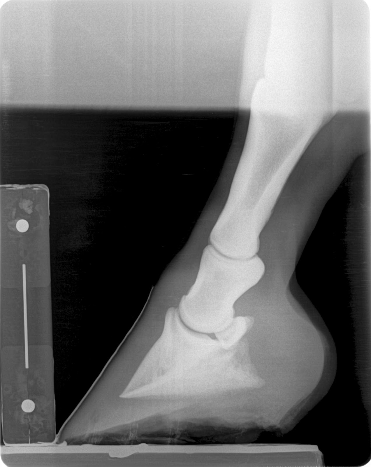
2. Balanced mechanical forces- This information is obtained from careful and in depth examination of external hoof characteristics combined with information based on measured soft tissue parameters from a farrier friendly radiograph. Radiographs must be taken in a consistent manner to obtain results that can be compared between radiographs. Radiographic measurements that are important to evaluate are: Coronary band/Extensor process distance (CE), proximal (top) and distal (bottom) horn lamellar zone (H/L), digital breakover (DB), sole depth (SD), and palmar angle (PA). Accurate assessment of these parameters will give you an idea if the horse's hoof is within a healthy range or not. To be successful in many common foot ailments, such as laminitis, navicular syndrome, caudal heel pain, long toe/low heel and club feet, it is paramount that precise radiographic evaluation of the forces at play is accomplished. The basis for all my therapeutic recommendations comes from these measurements. Below is a diagram of soft tissue parameters commonly utilized.
Farriers are often given a very vague prescription such as wedge the heels and back the toe up, but how much wedge and where should the toe be backed up to. A more precise prescription might include: DB at 0 mm, PA increased from 0 degrees to 10 degrees and use of aluminum rail shoe rockered mid shoe attached with glue and fit with a positive pressure frog plate. In order for a prescription like this to be given and received both farrier and veterinarian must speak and understand the same language, which also means that both individuals have pursued a higher level of understanding of the equine hoof.
3. Preventive hoof care programs- Many equine hoof ailments are results of long standing minor mechanical imbalances and predisposing genetic traits. Many of these can be identified early in life and monitored on annual basis via farrier friendly radiographs. For example, if your horse as a yearling has long pasterns, zero degree pa and a 30 mm breakover then you can assume that as an adult he will be predisposed to crushing his heels, maybe have thin walls and sole. However since it has been identified at an early age a maintenance program for the farrier can be developed that may differ from a basic perimeter fit steel shoe or traditional trimming. Many horses these days are not blessed with perfect feet and many would benefit from minor modifications in shoeing approach early in life to help delay or prevent the onset of hoof disease. A preventive hoof care program should involve a yearly podiatry style exam with radiographs that could be easily included into your yearly vaccination and wellness exam. Foals should be evaluated within the first week of life and every month for the first year of life. Radiographs can be taken any time along the way but definitely prior to entering training as to develop a hoof care plan. We as hoof care professionals need to be focused on maintaining hoof mass and quality instead of pretty and appealing to the eye. We can find minor changes in the measured soft tissue parameters long before bone changes occur and before the horse will exhibit pain or discomfort.
4. Regular and consistent farrier visits- It is very important to have shoeing/trimming intervals that are appropriate for the individual horse as mechanical properties and soft tissue parameters change early in the shoeing interval. Often times by the end of the shoeing period, especially if overdue, the soft tissue measurements such as palmar angle and digital breakover have entered into an unhealthy zone. Using the podiatry style radiograph to design a healthy protocol that may maintain a healthier palmar angle and digital breakover longer in the shoeing cycle is another added benefit for preventive hoof care programs.
In conclusion, I would like to see veterinarians and farriers alike adopt this similar language and radiographic techniques to evaluate the equine hoof. Without regard to consistent technique and a detailed evaluation of the mechanical formula there is an inherent risk of not obtaining the level of success that one may desire. What we do, and more importantly what we do not do to the hoof, not only has an affect today but in the future as well. We all need to recognize that a perimeter fit flat steel shoe may not be the best option for every horse, as simple modifications may prolong the health of the foot and prevent problems down the road.
Further reading and resources:
1. Dr. Redden's website, www.nanric.com, numerous articles regarding evaluation and treatment of many common foot ailments and soft tissue parameter measurement illustrations and articles.