Welcome, About us

Hello and welcome. My name is Sammy L. Pittman, DVM and I am a veterinarian, farrier, and horsemen with a great interest in the field of equine podiatry. My wife and I own and operate Innovative Equine Podiatry and Veterinary Services in Collinsville TX. My passion lies within the health and well being of the hoof to better serve your equine companion. With so much lameness attributed to the lower limb many horses require an out of the box approach to achieve the success desired.
Give us a call and we will be glad to help you in any way we can. Thanks so much.
I will be discussing different Cases and thoughts from our world with the horse. Feel free to contact us via text or call at 903-718-0056 or send an email to iepvs11@gmail.com. Thank you for reading and enjoy

Friday, October 26, 2012

Images from the October clinic with Dr. Ric Redden.


Hello,  I wanted to thank all the attendees to the in depth equine podiatry lecture and demo with Dr. Ric Redden of International Equine Podiatry Center.  We had some very good cases in which to apply a sound and methodical radiographic and external evaluation.  Using this evaluation a therapeutic shoe was apply to aid in rehabilitation of each case.  

We are planning a follow up clinic November 17th to recheck and reset each case.  This will be a great opportunity to see the response radiographically.  


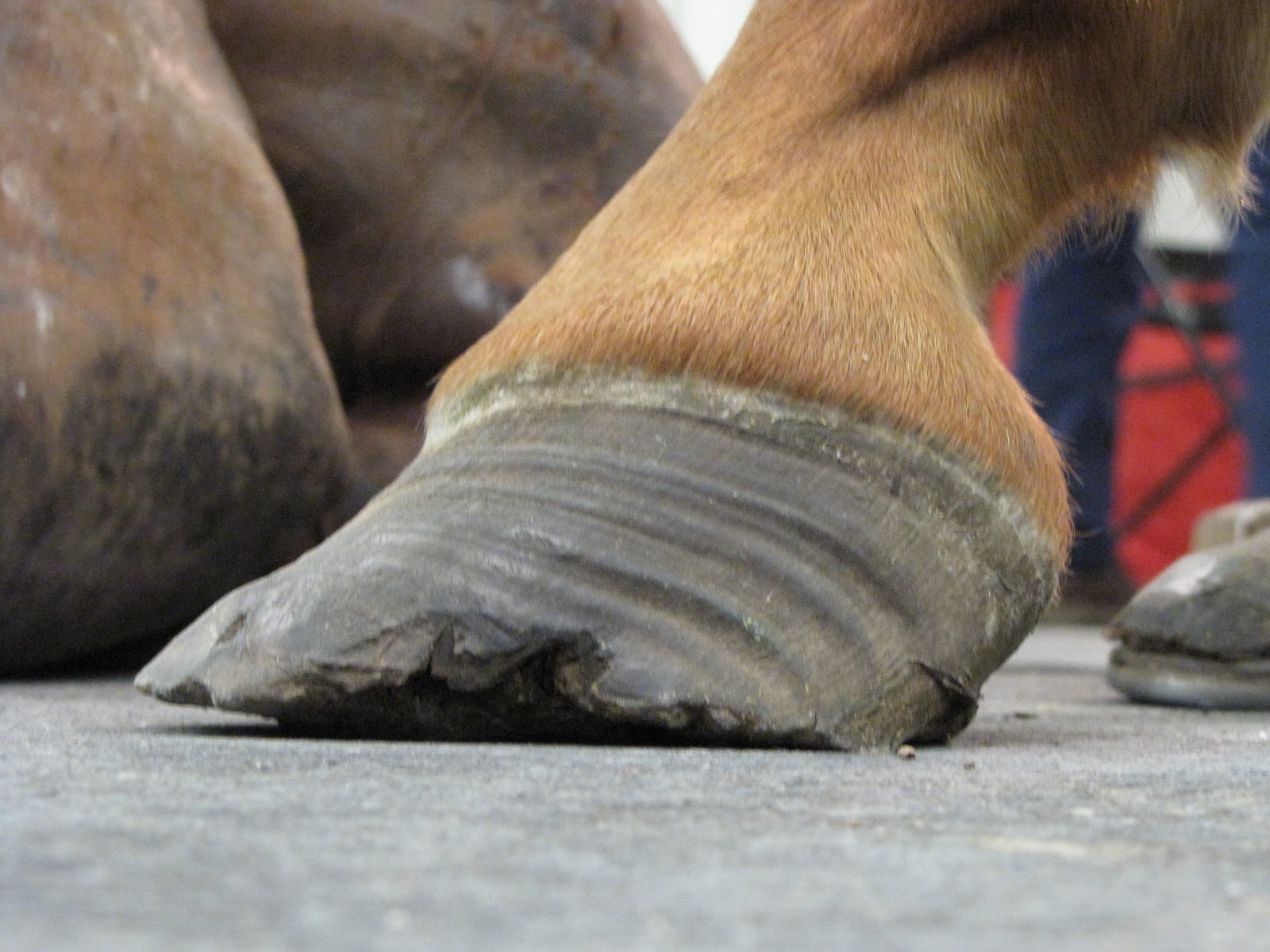
This first case is a grade 2+ club cases used as western and english pleasure as well as some roping.  


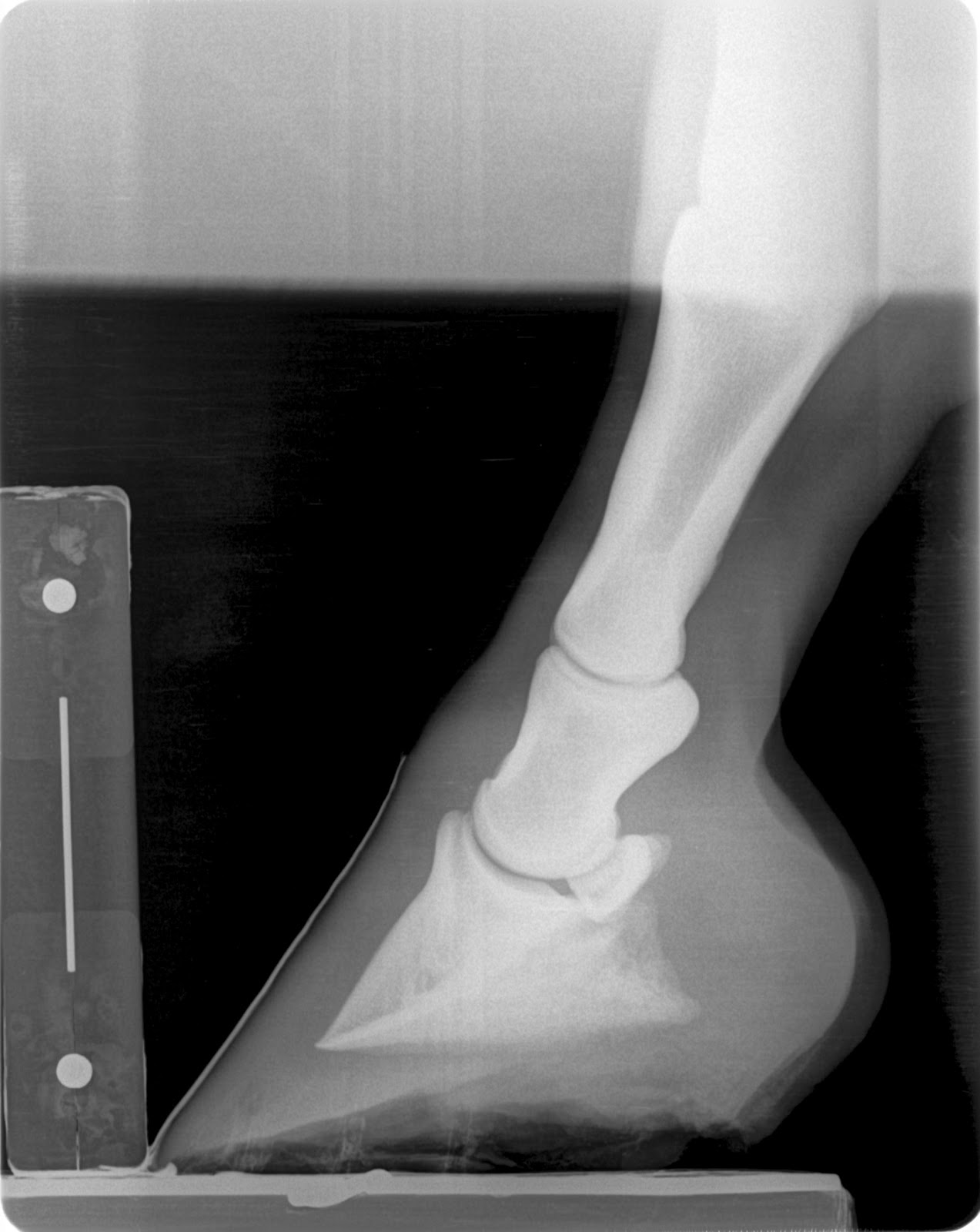
This is a case with navicular bone changes that had responded to increase in palmar angle and reduction in digital breakover but was not consistently going sound.  Dr. Redden applied an aluminum rocker rail.  Look at the TSA and the distance the navicular is from the proximal p2 between the pre and post shoe radiographs.


This patient has had some undiagnosed recurring lameness.  Today no in depth workup was performed but a shoe to enhance foot mass recovery and increase sole depth was applied.


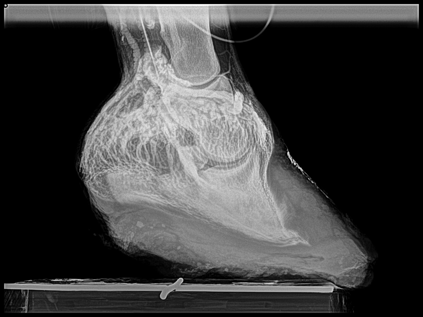
This is a many year chronic laminitis.  Goal with the rockered aluminum rail is to decrease DDFT tension unloading the apex of the coffin bone and the tension forces at the horn/lamellar zone.  This places breakover in the center of articulation.  This will improved compromised circulation in the dorsal region of hoof and a more even hoof growth from toe to heel is expected as well as improved sole depth.


This case had an acute bout of laminitis about 6 months ago.  Venograms show that circumflex is above the apex of  coffin bone and compressed tightly to tip of coffin bone as well.  Dorsal lamellar zone on the right front is broken and a void of contrast is present at coronary plexus.  No solar papillae are evident even at an increased to 20 degree palmar angle which should unload DDFT by 60 percent.  Treatment included derotational shoeing followed by a deep digital flexor tenotomy.


Monday, September 3, 2012

Cyril's Hoof Spring

This is a technique I learned from a farrier from Switzerland (Cyril Zuber) while at Dr. Redden's advanced equine podiatry class last month.  This case experienced a severe heel bulb laceration about a year ago.  All has healed up but a severe contraction secondary to loss of heel mass from injury and lack of full load bearing has occured.  I can't say if the contraction component is related to any lameness but a more cosmetic hoof could be obtained.

Below is radiographs and photos of the hoof and spring application process.  At the very end is a video of the spring being released showing the action that it is applying to the hoof capsule.

I placed in a custom steel rocker rail to fully load heels and maintain a self adjusting palmar angle with greatly reduced toe lever.


Friday, August 31, 2012

New navicular case

I have been too busy to post many cases in the last few months.  Thanks to Isaac we are getting some rain which allowed me a few hours on the computer

 The following is a case from this week.  This is a 7 year old  pleasure horse with a long term history of choppy gait and off and on head bob responsive to bute.

1/5 lame bilateral but 2/5 on left turn in a tight circle.  Left front is a grade 1+ club and podiatry style films confirm healthy soft tissue parameters.  My thought process is:  With healthy sole depth and minimal remodelling of the apex of coffin bone on a club foot I want to next look at the navicular bone to evaluate for lesions.  This case shows significant lesions in several views.  This are most likely in the distal half of the flexor cortex of the navicular bone.  I find these similar lesions in younger non lame patients but I do feel as they reach middle age they begin to create lameness.  Dr. Redden feels that the navicular bone of the club foot does not recieve as much load from the tendon versus the lower profile foot and does not develop as strongly as the low foot.  I am beginning to feel that these lesions are developmental and become more degenerative with age and use.  If dissected out these lesions will be a slight depression in the flexor cortex with a roughened edge that likely causes some abrasiveness to the deep digital flexor tendon.

My treatment plan is to drastically increase palmar angel to reduce load on the painful area.  This will allow some decrease in inflammation and likely a lower mechanical shoe in the future.  I have also prescribed a 5 day course of bute to decrease pain and inflammation.  I choose mechanical options first as it is a very low risk treatment and we can always resort to injections of the coffin joint and/or navicular bursa.  However I find that most of my cases respond very well to mechanical enhancement alone.  I will let you know about the response we obtain in this case.

Below are images of the navicular bone, pre and post shoe podiatry radiographs and digital photos of the patient.

Click here to shop Dr. Reddens products (shoes, ultimates and much much more) SHOP NANRIC


 Note the better digital alignment the drastic change in palmar angle, tendon surface angle and reduced toe lever.  This shoe changed PA by 12-14 degrees which will unload the tendons load on the navicular bone by 50 to 60 percent.


Thanks for looking.

Tuesday, July 24, 2012

Upcoming Clinic with Dr. Ric Redden

Come join us for a unique learning opportunity!  Hope to see you there.  If I can answer any questions regarding this lecture/demo please feel free to call 918.235.1529 or email at iepvs11@gmail.com