Welcome, About us

Hello and welcome. My name is Sammy L. Pittman, DVM and I am a veterinarian, farrier, and horsemen with a great interest in the field of equine podiatry. My wife and I own and operate Innovative Equine Podiatry and Veterinary Services in Collinsville TX. My passion lies within the health and well being of the hoof to better serve your equine companion. With so much lameness attributed to the lower limb many horses require an out of the box approach to achieve the success desired.
Give us a call and we will be glad to help you in any way we can. Thanks so much.
I will be discussing different Cases and thoughts from our world with the horse. Feel free to contact us via text or call at 903-718-0056 or send an email to iepvs11@gmail.com. Thank you for reading and enjoy

Monday, March 25, 2013

Acute laminitis case showing the value of the venogram


This is an acute laminitis case that was initially painful and exhibiting the typical founder stance on December 16 2012.  No known cause but a mild colic episode had occurred 3 days prior.  I was able to perform venograms on what I would consider Day one of laminitis.  These plain film radiographs and venograms create a baseline in which to compare follow up venograms.  This allows assesment of the mechanical therapy applied and severity of compromise in the days to follow.  Venograms 5 days later show moderate compromise already.  Consider the amount of compromise present while treated with mechanics. Unloading the Deep flexor tendon, its forces applied to the dorsal lamellar zone and sole directly below tip of coffin bone via raising the palmar angle.

There is no measurable displacement of the bone but significant change has occured within the vascular network as early as five days.  Some financial constraints existed and it was decided to wait 30 days and repeat venograms to evaluate if the mechanics applied would be enough.  I warned the client at this point that a deep flexor tenotomy may need to be performed as this rapid progression is very concerning.  Ideally a repeat venogram in another 4-7 days would have been best but finances limited to a 30 day check.  Knowing what I know now about this case I would have cut tendons on day five and never looked back.  Below is the Day one venogram on the left compared to the Day five Venograms on the right side.  Focus your eyes on the circuflex artery, terminal papillae and the vasculature directly below the tip of coffin bone.  Moderate compression and diplacement of these vessels five days into the syndrome.  Remember that no measurable rotation, increase in h/l zones, loss of sole depth has occured.


After applying the modified ultimates on day one the horse began to make a clinical improvement noted by moving around in stall better and better appetite.  He no longer rocked back to make turns.  This is important to consider as clinical signs alone are not good indicators of how the case is going.  The horse will lie to you when comes to laminitis.  


Right Front venogram comparson Day one to Day five
Left Front Venogram comparsion Day one to Day five.
Day 5
Day five


Below are venograms that are 30 days post laminitis of the left front.  Note the circumflex is several milimeters above the tip of the coffin bone and no papillae are present.  I again recommended deep flexor tenotomy as the modified ultimates are not providing enough mechanical relief, through unloading the force of the deep flexor.  The tenotomy abolishes all forces applied to the bone to hoof attachments and solar corium directly below tip of coffin bone.  Finances prohibited the Owner from going the surgical route and horse was actually improving with regard to pain and we decided to wait another thirty days.  

30 Days Post.  The Circumflex artery is displaced several mm above the tip of the coffin bone and the solar vasculature is tightly compressed below the tip of the coffin bone.
Below are 60 day post radiographs.  Note the increased C/E distance, diverging H/L zones (rotation),and loss of sole depth, especially on the Left.  No growth is noted on examination of the dorsal hoof wall and about 1/4 in at the heels is noted. Just to recap, we have no measurable displacement of the bone until 60 days into the syndrome but significant vascular changes on day five that continues to fail despite mechanical therapy.  If you are waiting to diagnose laminitis based on rotation you are 60 days late in this case.  

60 day post insult radiograph.  Note changes in ce, hl zones and sole depth.
60 Days post initial insult.  Note diverging hl zones (some may call rotation) increased in ce and comparable decrease in  sole depth.


Below are comparative venograms of the left front.  On the left is day one and on the right is 90 days after intial insult.  Note the greatly altered circumflex architecture and the tip of the coffin bone is visualized below the solar vessels.  Moderate thickening of the dorsal sublamellar zone and significant accumalation of contrast  as the dorsal coronary band that is likely secondary to a septic process that is brewing.  Horse still has not grown any sole or dorsal hoof wall in 90 days.  Note the scallop like loss of bone that occurs just above the tip caused by displacement of the terminal papillae and circumflex artery.  I can only assume the combination of local hydraulic pressure and possibly the redirected papillae in this area are to blame for this unique change in the shape.  At this stage in the game many irreversible changes have occurred that will likely prevent this horse from returning to a previous level of competition.  
90 days post comparison

90 days post comparison .  The right foot has suffered less but prominent and significant change/damage has occurred in the area of the circumflex artery and terminal papillae.
Left front Post tenotomy and derotation.  

Right Front post tenotomy and derotation.
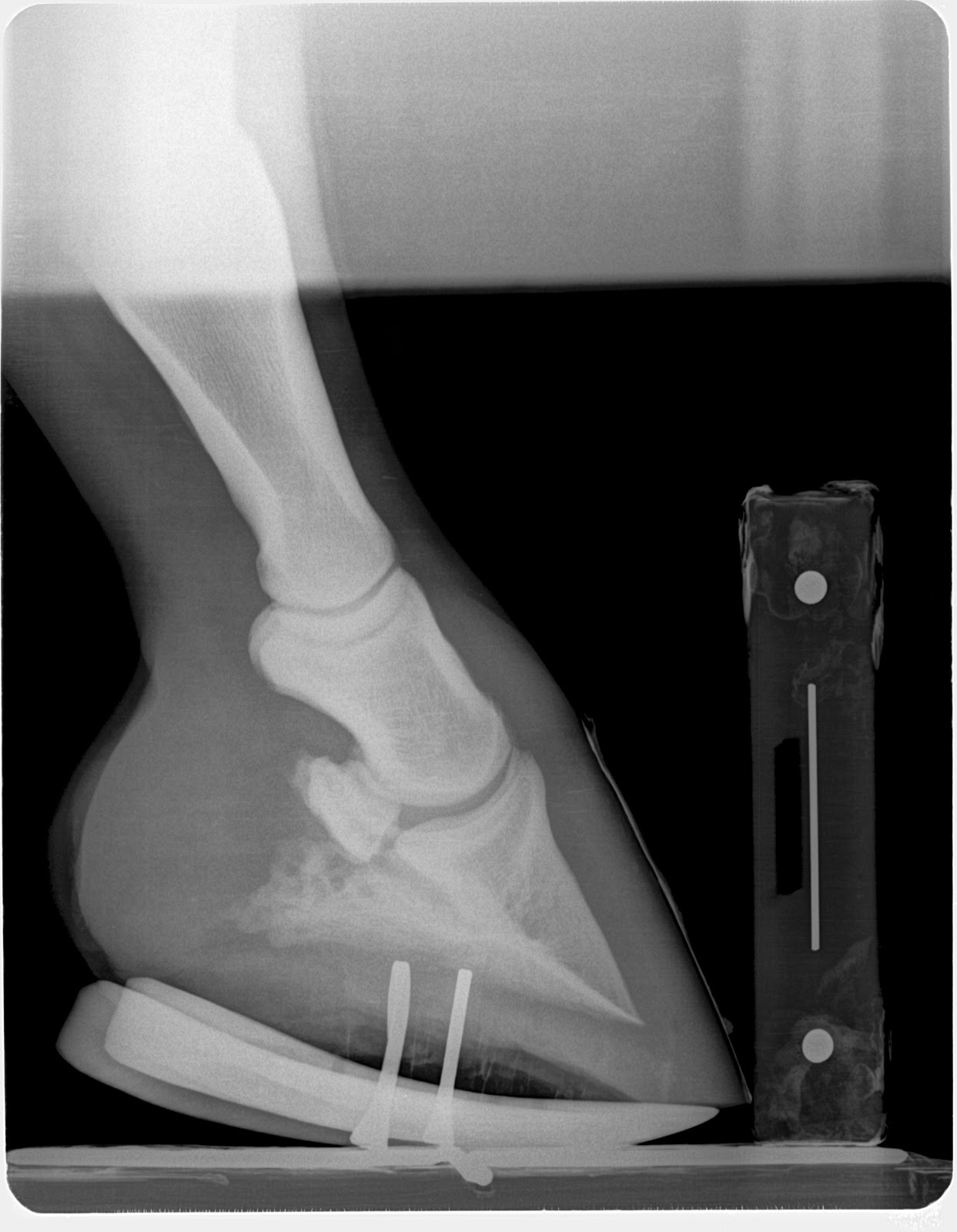
At the 90 day examination I advised the client that surgery was the only option for treatment and this late in the game I could only offer a guarded prognosis for any return to previous level of performance.  The owner opted to donate the horse for a learning seminar.  On March 22, 2012 a little over 90 days post initial insult a eager bunch of vet students and farriers performed venograms, applied derotation shoes and performed deep flexor tenotomies.  In the days following the horse is moving much better.  I will post followup radiographs in 30 days to evaluate the progress.  I do feel recovery is still very likely.  Ideally performing the tenotomy prior to irreversible damage and bone change would result in a better outcome.


In conclusion I would like to re iterate the important points:
1)  In hindsight a deep flexor tenotomy should have been performed on day five or very shortly after.
2)  Improvement with regards to pain and movement are not good indicators of success in laminitis.
3)  Radiographic measurements alone, early in the syndrome, may change very little if any and a venogram may be the only information alluding to the nature of the insult.
4)  Aggressive early mechanical therapy did relieve a lot of pain but did not provide enough unloading of the Deep digital flexor to aid in unloading of the vascular supply and further unloading via deep flexor tenotomy would have likely given a better outcome if performed earlier.
Stay tuned!

Tuesday, February 5, 2013

My Take on barefoot management


I get this question alot and I thought it would be a good question to address.  Let me start by saying I love managing horses barefooted and it would be wonderful if that is all it required to fix many hoof problems.  The truth of the matter is that many horses need shoes to alter the mechanics to their advantage to maintain good sole depth and soundness.  Many of today's horses have what I call genetic handicaps that prevent them from have the ideal foot and forces applied to it.  Horses that can stay sound barefoot and compete will have the following parameters measured radiographically:  sole depth of 15mm, bone angle 50 degrees, a positive palmar angle, a toe lever (measured from center of coffin joint articulation to the tip of coffin bone) that usually is not much more than 60 percent of overall coffin bone length, growth that is equal to or exceeds 7-10mm in 45 days and is even from to heel.

Change a few of these parameters and you start to identify mechanical handicaps that increase forces on growth centers and reduce blood flow hence reducing horn/sole growth.  The most relevant difference I see is the bone angle,  the toe lever and relative deep digital flexor tension/length.  These are characteristics that a horse is born with.  A lower bone angle and longer coffin bone (toe lever) often go hand in hand.  This creates a longer lever arm acting against the flexor tendon and the bone to hoof attachment at the toe.  On the other hand take a club foot with shortened muscle/tendon unit that overloads the toe area and this excessive force is responsible for the lack proper circulation and growth even with a shorter toe lever.  Depending on the severity of these genetic handicaps and the goal for the horse will determine whether or not   your horse will do good as a barefoot candidate.  
Note a short toe lever.  When evaluated from COA to tip of coffin bone.  This horse maintained good sole depth and hoof quality with minimal trimming.  

Note the long Toe Lever when evaluated from COA to tip of coffin bone.  This horse would likely benefit from a shoe package that reduced the toe lever (rolled, rocker toe, rocker shoe, natural balance style)  as an maintanence of genetic handicaps.  This case was place in rocker rails for initial management then to a rockered keg shoe.  


Yes, sometimes a shoe can be detrimental to the hoof if attention is not given to recognizing these handicaps and altering the mechanics with the shoe application.  For example a long toe lever and long coffin bone would benefit from a shoe application that reduced the toe lever.  Examples are a rolled toe, rocker toe, natural balance or rocker shoe.  This allows a reduction in the moment arm and reduces the force transmitted against the deep flexor tendon. This reduces subsequent tension force on the horn-lamellar junction, compression force at the solar corium and navicular bone tendon interface.  

I feel that earlier recognition  and management of the these handicaps will reduce many problems that our horses experience later in life.  Horses are such amazing beast and able to adapt and overcome problems for many years to only be painful in their later years.  I feel a reduction in these forces, once recognized and shoeing plan altered, prior to training would reduce the accumulated trauma over the lifespan of the horse.  

So my overall stance is:  If your horse has good external and radiographic parameters and no genetic handicaps or disease processes that require unloading then by all means manage with a good barefoot trim. However if there is problem such as thin soles, navicular bone disease, laminitis, arthritis or white line disease, application of a shoe to place the forces in the horse's favor may be required.  Many cases require our modification of the forces to keep the horse doing its job.  

I think shoeing horses often gets a bad reputation because we fail to recognize that maybe a perimeter fit, flat shoe may not be the best for every horse.  I see many that would be better off  barefoot as the shoe is creating an ever lengthening toe lever that may have the chance to wear off, if the horse was barefoot.  

This horse is nine years old and have never been trimmed.  Note the  load zones.  This horse  is maintaining his own four point trim.  Not that no hoof care is good but this horse has a foot that is sound enough not only to go barefoot but has the right environment and proper mechanics to maintain itself.


So when and if I can manage horses barefoot I prefer the four point trim.  It's ability to reduce digital breakover greatly enhances the quality of and quantity of growth.  I think the secret is "less is more"  This approach allows maintaining a short toe and toe lever with maximizing sole depth.  This trim is typically a rasp only trim with minimal knifing of the frog and sole only to clean up any ragged frog or bars that may trap debri.   Below is a short video demonstrating a four point trim and some images demonstrating some of the important aspects as well.  


Points to remember:

1) Very good trimming approach to manage horse barefoot even with long toes and club as long as there is no other problems
2) Get paid for what you leave on the horse not what you are taking off.  
3)  When transitioning from shoes to barefoot, Take baby steps in applying any trim unless you have a ton of foot.  Consider trimming every 2-3 weeks for 3 trims until you get the foot toughened. 
4) I see great benefit from the use of Keratex hoof hardener and hot searing (Propane torch) my barefoot cases
5) Leave a nice round radius to the hoof wall and the hoof wall will be less likely to chip and more likely to self maintain.
Angle of Rasp for toe trim varies according to sole depth.

Demonstration of the load zones with a four point trim


Sunday, January 13, 2013

The grey are aka "the hoof" article


The Grey area aka the hoof


            As horse owners, farriers, trainers and vets we all know about the ever increasing foot ailments that horse's endure.  We have all heard the saying, “No foot, No horse”.  Do we really live that approach in our day to day lives with our equine companions?  Have we really obtained all the information possible about our horse's hoof dynamics?  The majority of hoof lameness' and even upper limb lameness' are a mechanical diseases that can benefit from a well developed mechanical solution based on evaluation of the forces at play within the hoof combined with accurate diagnosis and medical therapy. The hoof is often times overlooked as many of the people involved in the care of horses do not have all the information necessary to help maintain a healthy foot.  Farriers have good working knowledge of trimming, nailing, using various tools in there day to day job but many lack knowledge of internal anatomy, radiographic anatomy and physiology.  Veterinarians have a good understanding of anatomy, physiology and diseases but lack many of the hands on skills,  knowledge of external hoof characteristics and techniques that a farrier takes for granted.    The grey area is birthed from neither profession has enough information to communicate on the same level.  As a veterinarian I was not educated on bio-mechanics, how to take farrier friendly radiographs, or how to evaluate lower limb mechanical forces.  There just isn't enough time to completely cover all aspects of the horse while in veterinary school. Most veterinarians base their therapeutic recommendations on findings in veterinary lameness text or based on empirical personal experience and not a well designed mechanical plan based on radiographic findings.  I know this because that was my approach upon graduating veterinary school.  I find in my everyday practice that many hoof care professionals are unaware of a more in depth approach to evaluating and treating hoof disease and lameness.  When we combine the knowledge of both professions with egos aside and develop a plan from that combination more success will arise.  Many foot ailments can be a financial and emotional drain and require aggressive, quick and precise mechanical and medical treatments to be successful.  I have been fortunate to learn from a pioneer in the podiatry world, Dr. Ric Redden of Versailles, Ky.  Through his practical and innovative use of venograms, serial podiatry style radiographs and new mechanical devices, many horses are relieved of unnecessary pain and suffering. 

            Below are four basic guidelines for successfully maintaining healthy hooves and approaching hoof lameness issues. 
1.     

Nutrition- We are all aware that skinny horses do not typically grow good hooves, but did you know that research has shown that added biotin at a rate of 100mg per day will increase hoof quality.  Common hoof supplements that are commercially available only supply 10-20 mg daily. Biotin is long been noted to aid in hair and hoof growth.  All of my hoof cases that have poor quality hoof,  thin soles, slow growth or laminitis (founder) are started on 100 mg of Biotin daily.
2.      Balanced mechanical forces-  This information is obtained from careful and in depth examination of external hoof characteristics combined with information based on measured soft tissue parameters from a farrier friendly radiograph.  Radiographs must be taken in a consistent manner to obtain results that can be compared between radiographs. Radiographic measurements that are important to evaluate are:  Coronary band/Extensor process distance (CE), proximal (top) and distal (bottom) horn lamellar zone (H/L), digital breakover (DB), sole depth (SD), and palmar angle (PA).  Accurate assessment of these parameters will give you an idea if the horse's hoof is within a healthy range  or not.  To be successful in many common foot ailments, such as laminitis, navicular syndrome, caudal heel pain, long toe/low heel and club feet, it is paramount that precise radiographic evaluation of the forces at play is accomplished.  The basis for all my therapeutic recommendations comes from these measurements.  Below is a diagram of soft tissue parameters commonly utilized.      
                                                                                                                                                                                                                                                                                                                                                                                                                                                                                                                                            Farriers are often given a very vague prescription such as wedge the heels and back the toe up, but how much wedge and where should the toe be backed up to.  A more precise prescription might include:  DB at 0 mm, PA increased from 0 degrees to 10 degrees and use of aluminum rail shoe rockered mid shoe attached with glue and fit with a positive pressure frog plate.  In order for a prescription like this to be given and received both farrier and veterinarian must speak and understand the same language, which also means that both individuals have pursued a higher level of understanding of the equine hoof. 
3.      Preventive hoof care programs-  Many equine hoof ailments are results of long standing minor mechanical imbalances and predisposing genetic traits. Many of these can be identified early in life and monitored on annual basis via farrier friendly radiographs.  For example, if your horse as a yearling has long pasterns, zero degree pa and a 30 mm breakover then you can assume that as an adult he will be predisposed to crushing his heels, maybe have thin walls and sole.  However since it has been identified at an early age a maintenance program for the farrier can be developed that may differ from a basic perimeter fit steel shoe or traditional trimming. Many horses these days are not blessed with perfect feet and many would benefit from minor modifications in shoeing approach early in life to help delay or prevent the onset of hoof disease.  A preventive hoof care program should involve a yearly podiatry style exam with radiographs that could be easily included into your yearly vaccination and wellness exam.   Foals should be evaluated within the first week of life and every month for the first year of life.  Radiographs can be taken any time along the way but definitely prior to entering training as to develop a hoof care plan.  We as hoof care professionals need to be focused on maintaining hoof mass and quality instead of pretty and appealing to the eye.   We can find minor changes in the measured soft tissue  parameters long before bone changes occur and before the horse will exhibit pain or discomfort. 
4.      Regular and consistent farrier visits-  It is very important to have shoeing/trimming intervals that are appropriate for the  individual horse as mechanical properties and soft tissue parameters change early in the shoeing interval.  Often times by the end of the shoeing period, especially if overdue, the soft tissue measurements such as palmar angle and digital breakover have entered into an unhealthy zone.  Using the podiatry style radiograph to design a healthy protocol that may maintain a healthier palmar angle and digital breakover longer in the shoeing cycle is another added benefit for preventive hoof care programs.
           
            In conclusion, I would like to see veterinarians and farriers alike adopt this similar language and radiographic techniques to evaluate the equine hoof. Without regard to consistent technique and  a detailed evaluation of the mechanical formula there is an inherent risk of not obtaining the level of success that one may desire.   What we do, and more importantly what we do not do to the hoof, not only has an affect today but in the future as well.  We all need to recognize that a perimeter fit flat steel shoe may not be the best option for every horse, as simple modifications may prolong the health of the foot and prevent problems down the road. 

Further reading and resources: 
1.      Dr. Redden's website, www.nanric.com, numerous articles regarding evaluation and treatment of many common foot ailments and soft tissue parameter measurement illustrations and articles.

Theory of two major loads article


Theory of Two Loads


            I have struggled with what forces are involved in the hoof and how they changed with different palmar angles and varying degrees of deep digital flexor tension (DDF). So to aid in my understanding I consider two extreme examples to help describe my simplified idea of two major loads within the hoof capsule.  First I will describe tendon load (TL) and the extreme example to be used is a high grade club.  Next we will discuss bone load (BL) or ram load with the extreme example of a post ddf tenotomy laminitis case. 
             I think we can all agree that there is a significant pull from the DDF in club foot cases.  Lets consider the action of the DDF.  As weight is applied to the limb or the DDF muscle contracts the  pulling force is transferred to the coffin bone via the semilunar crest at the DDF tendon insertion.  This pulls the coffin bone around its articulation with the distal end of the second phanlanx (P2) and the DDF tendon also is pressed against the flexor surface of the navicular bone.  Extraction forces are apparent at the horn-lamellar interdigitation and compression forces on the solar corium directly beneath the apex of the coffin bone.  Club feet are affected by a shortened musculotendonous unit via increased neurologic stimulation of the flexor muscle.  This tranfers load to the apex of the coffin bone and the horn-lamellar interface at the toe.  So for simplicity sake consider two lengths of rope both attached above carpus and at the semilunar crest of coffin bone.  The shorter length will transfer more load to the apex than the longer when weight is applied to the limb.


                                              Figure 1 short rope/high pa/club
                                                         
                                            Figure 2 Longer Rope/low pa/slam dunk


These forces and the changes implied are noted on radiograph's of club feet, as a remodeled tip of coffin bone, a small bump midway down on the face of P3 and often smaller,and a less dense navicular bone.  These changes follow Wolfes law of bone remodels along lines of tension and compression.  Now consider the external characteristics of this extreme example:  Atrophied frog, deep central sulcus, wider growth rings at heel than toe, bulging or flat sole at and around apex of frog.  These characteristics are created by the excessive DDF tension which allows for an unbalanced load distribution between tendon load and bone load.  This excessive TL prevents loading and stimulation of the palmar portion of the hoof and leaves the frog and heel suspended in the air.

.              
Figure 3 bone remodeling on tip of coffin bone


            The second load to consider is bone or ram load (BL).  This is the weight that is transferred through the bony column directly to the ground.  If no DDF was present then all load is distributed through this manner and forces are increased in the heel region.  Consider the case of a post deep digital flexor tenotomy when all TL has been negated due to severing of the   DDF tendon. All weight and forces are concentrated in the heel region and has more of a table leg distribution of forces.  I feel that many of the crushed heel, low to negative palmar angle hooves  have a similar situation. Just as the club foot is born with shortened musculotendinous unit the low Palmar angle/crushed heel or slam dunk foot may have a longer than ideal musculotendinous unit allowing a greater bone load that will allow more weight or load through the bony column to the palmar/plantar aspect.  I think it is possible to create a negative palmar angle and crushed heels with poor mechanics in many of our everyday shoeing practice that could possibly take a normal healthy foot with good sole depth and palmar angle to thin soles and negative palmar angle, however many are destined for that path from a very early age due to conformation.  It is impossible to take a  club foot caused by shortened musculotendinous unit and create a negative palmar angle and the same may be true for the slam dunk foot as many will revert back to crushed and under run heels once orthotic devices have been applied to increase hoof quality, sole depth and aid in treatment of lameness. 
         Consider a heel sore horse that is landing toe first, this is evidence to me that the horse can use the tendon to transfer load to the front of the foot to unload the painful buttress, digital cushion and many related soft tissue structures.  Many horses compensate quite well by transferring load to the front of the foot via DDF with initial heel soreness but it is not long until the extra workload by the tendon creates inflammation within the tendon itself  and many of soft tissues and ligaments associated with the palmar/plantar aspect and fatiguing the flexor muscle group.  This is when a trip to the vet usually occurs as they are now unable to effectively transfer load to a non painful region and show obvious signs of lameness.  The increased load transferred to the front by the toe first landing and often long digital breakover in these cases decreases blood supply to vital growth centers and adds to the further compromise of hoof and sole quantity and quality.  Radiographs would show very thin soles below wings of coffin bone, low to negative palmar angle, a very low tendon surface angle, as scallop of bone remodeling in palmar/plantar aspect of solar margin of coffin bone and upright pasterns.  External characteristics noted are:  Wider growth rings at toe than heel, flat and thin soles, 2-3 sets of nail holes, wide robust frog, and under run heels.


                                     Figure 4Low Pa bone remodeling/low ddft tension
           
For further understanding let us consider treatment of these two scenarios and why they are successful            l in increasing soundness and quality of hoof mass.  For the club foot syndrome, lower grades that are not surgical candidates, increasing palmar angle and lengthening the heel base will allow more BL and less TL.  Decreasing the TL will decrease the amount of load being transferred to the toe and allow more bone or ram load to push into the heels.  The easiest and most successful approach I have found, is using rocker shoe mechanics.  The heels are trimmed to the widest part of the frog parallel to the wings of the coffin bone and toe is trimmed perpendicular to the frog axis at a low rocker toe style angle.   
                                    
                                                                 Figure 5 Grade 3 club
    
              Figure 6 Grade 3 club with Rocker Rail

 The trim will vary based on such parameters as palmar angle, sole depth and digital breakover but the basic approach will stay the same.  The next step is to determine what shoe to shape to fit our specific needs.  In general low grade clubs will do fine in a rockered flat shoe as higher grade clubs may require starting with a wedged shoe that has greater mechanical potential.  Consider a flat shoe that is rockered can alter pa 2-4 degrees and a 5 degree rail shoe is starting with 5 degrees, so any added rocker will increase potential to alter palmar angle.  So the question to be answered is how much PA increase do I need to create less tendon load and more bone load?  Low grade clubs require less than higher grades.  This approach will allow more ram or bone load, more heel loading that will result in less atrophy of the frog, decreasing depth of the central sulcus, increased sole depth below the tip of coffin bone and more even toe to heel growth patterns.  With less TL comes less H/L zone extraction force and less solar corium compression. 

Now consider a case of acute lamintis with extensive H/L detachment and venogram shows decreased perfusion at the coronary waterfall, compromised vasculature down face of the coffin bone,  tip of coffin bone has displaced 3 mm below the circumflex artery, and terminal papillae are horizontal versus being in normal orientation with the face of p3.  This gives us a picture of severely compromised dorsal portion, including the horn-lamellar attachment and solar corium below the tip of coffin bone.  A DDF tenotomy may be indicated in many cases such as this. This will completely unload the forces of the DDF and allow all weight to be transferred down through the bony column into the palmar/plantar region of the foot and unloading much of the compromised areas in dorsal aspect.    This can be shown by post tenotomy radiographs and venograms.    This release and increased load now through the bony column to heels will often push the coffin bone up closer to its original placement prior to laminitis episode and displacement and radiographs will show measurable decrease in distal h/l zone and increase in sole depth just from the unloading that occurs from complete release of DDF.

                          Figure 7 laminitis with rotation


Figure Post tendon cutting and derotation shoeing  


Figure 9Acute laminitis venogram


 In the above drawings (Figure 7 and 8) shows the pull of the tendon with detached bone to horn attachments and a post tenotomy with derotational shoeing.  Without a healthy lamellar attachment there is no antagonistic force to counteract the pull of the ddft (TL) and the coffin bone rotates around its articulation compressing solar corium at the tip of the coffin bone.  Figure 9 shows an acute laminitis case in which the bone is compressing the blood supply at the tip of coffin bone due to lamellar detachment.  You can see the tip of coffin bone below the circumflex artery.  This area is heavily loaded secondary to the TL and loss of the bone to horn attachment.  The image on the right is of the same horse 2 weeks after derotational shoeing and deep digital flexor tenotomy.  The tenotomy negates all TL and its forces applied to the damaged areas (lamellar zone, sole under tip of p3) and heavily loads the palmar/plantar aspect of the hoof through BL only.  Note the restructuring of the blood vessels under and around the tip of the coffin bone in this short 2 week period. 
I have always considered that anytime we raise the palmar angle via wedges or rocker shoe mechanics that we increased the load on the heels but it really wasn't clear why until considering these two loads.  These examples are two extreme versions and most feet will fall somewhere in between.  When a healthy balance between TL and BL exist we find good  feet that are easy to maintain with adequate sole depth and a positive palmar angle but when loads sway more to one side of spectrum to overloaded portions become unhealthy and need our assistance in balancing the load via a well designed protocol based on and monitored by serial podiatry style radiographs and venograms.

Wednesday, December 5, 2012

Ric Redden, DVM follow up clinic 6wks rechecks

We had a great day resetting and rechecking many of the cases we used as demo's during Dr. Ric Redden's in depth equine podiatry lecture and demonstration.  We had some return students and some new ones attending.  I want to thank Clyde Brown and Animal Health Supply for allowing us to congregate at their place of business.

 Below is several follow up images and short discussion of each case.  Also look back at the previous blog entry for initial images and therapeutic shoe applied.  October clinic images link


White line disease Case:  Sole depth improved by 4mm but white line lesion failed to grow down at same rate and decision was made to remove hoof wall to expose oxygen and allow cleaning.  Owner reports that he is running around like a youngster again and is more comfy than is has been in a long time.
6wks post intial rocker rail note 4mm increase in sole depth in a horse that hasn't grown any sole in years.
Reset image
First image Oct 6 pre shoe


Hoof wall resection to allow cleaning and oxygen to penetrate


Club foot case:  This horse lost the rocker rail shoe applie to the foot opposite the grade 3 club (which is also a club) and regular farrier applied a flat steel keg shoe to keep foot protected.  Note the horn lamellar zone divergence.  One could call this rotation which would be non specific.  The divergence is created by the club syndrome stretching to lower horn to bone attachments.  This is confirmed by evaluating the dermal-epidermal junction and measuring the horn zone compared to the lamellar zone.  If the lamellar zone was larger than the horn zone one could conclude a laminitis as this is lamellar swelling.  In this case it is chronic stretching of the lamellar bone secondary to the constant pull of the deep digital flexor unit.

The Grade 3 club grew more sole in the rocker rail than did the lower grade club in a flat shoe.  This information tells us that placing the tendon sling in freedom with the rocker shoe allows better nutrient and blood circulation through unloading of the sole via reduced deep flexor tension.  We placed the grade 2 club (Left Front) in a rockered trim with rockered steel keg shoe to also place the tendon sling in release.  We will be to see a more rapid sole mass recovery in this hoof as well at the next reset.  Owner reports excellent comfort and has adjusted very well to the new shoeing approach.


Pre shoe radiograph Oct 5

Left front shoe that regular farrier had replaced with flat keg shoe for protection
Rockered keg shoe


6 wks post rocker rail application additional 4mm of sole and cup starting to form.  All this due to unloading of the deep flexor pull 


Chronic Lamintis case:  Farrier was a student and he reports horse is moving very nice.  Horse was able to stand comfortably for each shoe reset.  Turning and moving very nicely.


Pre Rocker shoe oct 6

6 weeks post rocker rail with addition of 4mm of sole and less bulge of sole at apex of frog.  
Oct 6th pre rocker 

Left front 6wks post rocker rail.  Rocker shoe was removed prior to getting a radiograph.  Added 5mm of sole

Nov 17th reset with rocker rail.  

Post nov 17th reset rocker rail.


Navicular case:  Owner reports she was able to work a pattern for the first time in 2 years.  The Owners farrier was present and we helped him reset the rocker rails.  We plan to maintian the higher palmar angle for the next shoe cycle then began to lower the mechanics/palmar angle.  I expect to achieve similar comfort with lower mechanics as the horse remained comfortable even with losing a few degrees of palmar angle secondary to growth.  The history is very important here.  If horse became more lame at the end of the cycle as the palmar angle decreased, this tells us the hot spot becomes loaded at the lower palmar angle and may require a longer period of higher mechanics.
RF pre reset on nov17th

Post shoe nov 17th

Post shoe nov 17th
Pre shoe reset on nov 17th


6 month chronic laminitis case:  Owner reports horse is very comfortable, has a much better appetite and very willing to move freely.  This case demonstrates the importance the deep digital flexor tendon force applied to a failed lamellar bone.  With the loss of the lamellar suspension of the coffin bone, it is allowed to compress the sole at the apex of coffin.  No blood, No growth and recurrent abscessation as has occurred in this case. The fragile rim of the coffin bone becomes loses its blood supply and acts like a foreign body.  I haven't been able to achieve this level of success with any other approach.  Doubling sole depth from 10mm to 20 mm in a matter of 6 wks in chronic laminitis is astonishing. 

I do not recommend a tenotomy for every laminitis case and only do so if the venogram shows the circumflex artery at or above the level of the tip of the coffin bone as described by Dr. Ric Redden.  However I do recommend considering the forces applied by the ddft to the coffin bone and often use "mechanics" (rockering/wedging) to lesson the tension on a failing system to aid in re-establishing vascular supply.

Immediately post derotation and deep flexor tenotomy oct 6


Note the rapid growth of sole at dorsal portion of hoof and loss of palmar angle.  addition of 10mm of sole

Post reset to re establish a zero palmar angle with the shoe.  This is necessary to prevent over correction resulting in a negative palmar angle 
Immediately post derotation and deep flexor tenotomy on oct 6

6wks post derotational shoeing and deep flexor tenotomy.  No reset required as even sole growth is occuring and resetting the shoe does not add any benefits mechanically.