


Come join us for a unique learning opportunity! Hope to see you there. If I can answer any questions regarding this lecture/demo please feel free to call 918.235.1529 or email at iepvs11@gmail.com

Open to all farriers, horse owners, trainers and veterinarians. Come join us for a review of gross and radiographic anatomy. Learn how to establish an appreciation for load dynamics based on radiographic and external characteristics. FREE!
I hope all are having a great holiday season. Kellee and I wish all of you the best in the new year. We are looking forward to new podiatry cases and meeting new clients.
I am finally getting some time to post some images from the Redden clinic we hosted here in Tulsa Ok back in October. We had a great time and had some good cases to work on. Two cases I have continued to follow with radiographs and one with venograms. Dolly the mild laminitis case we used as a venogram demonstration case I plan to post separately as a single case report after our next shoeing and venogram next month. She was an interesting case and I have lots of images. She is doing well and growing nice foot mass.
I can't thank Dr. Redden enough for spending his time with us in Tulsa. Anyone that may be interested in attending future clinics in Tulsa please email us a iepvs11@gmail.com.
I will also be posting an update to Lobo the laminitis case with recent radiographs, shoeing approach and venograms.
Please look back in the archives to view many other cases and images. I will be posting two more laminitis cases soon as well.
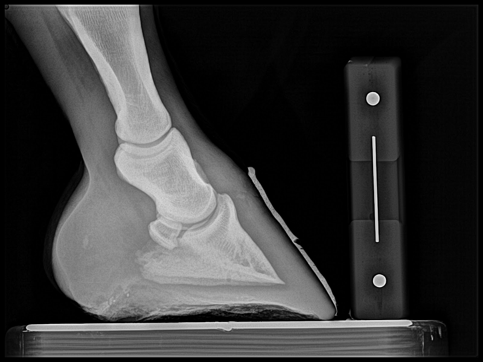
First case is a chronic navicular with rotational and varus limb deformities. Goal is to increase tendon surface angle, Palmar angle and reduce digital breakover.


Below: Quarter race horse that goes off after 150 yards or so and quits running. 4yrs old and has moderate navicular changes already. Placed in Rockered Race shoe from NANRIC to increase TSA and PA. This will unload DDF/nav bone engagement and small osteophyte at dorsal aspect of coffing joint.


Next Case: Older teenage trail and lesson horse. He has been plagued with Navicular disease and is 2/5 lame on left front. Immediate improvement is noted and absolutely no head bobbing at next reset. The first three images are from the day of the clinic and the last two are pre shoeing radiographs from the reset. I misplaced the day of the clinic rads for this case.


 |
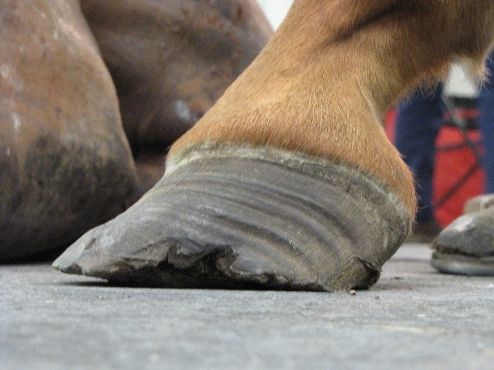
| This is is post trim with application of barium paste to accentuate the trim performed by Dr. Redden. |


 |
| Post 4 point trim. Note improved digital breakover created by the slight bevel in toe |
We had a great time, met some great people and had some great cases to work on. I felt there were several ah ha moments and several light bulbs came on during this clinic. I would like to thank Mr. Ellis for allowing us to have this clinic at his place, Amy Griffith for organizing and many others who helped. We had a little lecture time first thing on Saturday and Sunday morning covering basics radiographic soft tissue parameters that help determine the mechanical equation. A couple of venograms were performed to evaluate the vascular supply of different foot ailments. I will post some photos and radiographs of a few cases below. If you are reading this and you do not understand the purpose of evaluating soft tissue parameters and have not realized the benefit of the venogram please do not hesitate to ask questions. You can always email me at innovativeequinepodiatry@hotmail.com or post a comment here.
This is a Grade 3+ club. Notice the heels are not touching the ground after trimmed, the severe dishing of the dorsal wall. Both of these characteristics give indication of the level of Deep Digital flexor Tendon pull associated with club feet. If we accommodate the pull completely and not antagonize in any way this foot will be happy, as it will maintain plenty of sole depth and the dish will grow out. However, it will always require similar level of mechanical release or the dish will return and sole depth will decrease under the heavy pull of the DDFT. The rocker shoe allows the hoof wall at the heels to be trimmed to the widest part of frog which lengthens the load bearing surface and allows more loading of the heels. The rocker-ed surface allows the force of DDFT to rock up and not pull on dorsal (front) laminar attachments and compress the solar corium under the tip of the coffin bone. This unloading will allow rapid sole depth recovery.


 |
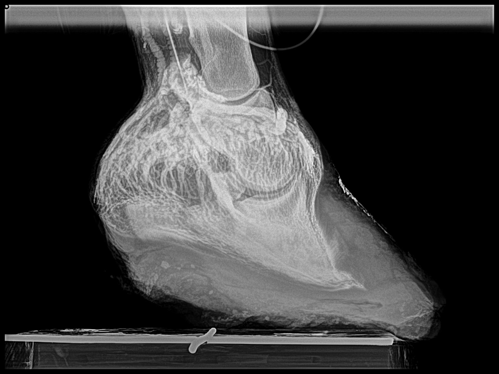
| Chronic lamintis with extreme Digital breakover |


Thanks again for everyones participation and I hope you enjoyed the hand's on experience. I hope you all have a great weekend. The Breeders invitational cutting horse show is in Tulsa town this week and we are excited to see some family and friends and wish them a good show. Thanks again for checking out my blog and hope to see you soon.
All the Best Sammy
Hello, I wanted to thank all the attendees to the in depth equine podiatry lecture and demo with Dr. Ric Redden of International Equine Podiatry Center. We had some very good cases in which to apply a sound and methodical radiographic and external evaluation. Using this evaluation a therapeutic shoe was apply to aid in rehabilitation of each case.
We are planning a follow up clinic November 17th to recheck and reset each case. This will be a great opportunity to see the response radiographically.
This first case is a grade 2+ club cases used as western and english pleasure as well as some roping. 


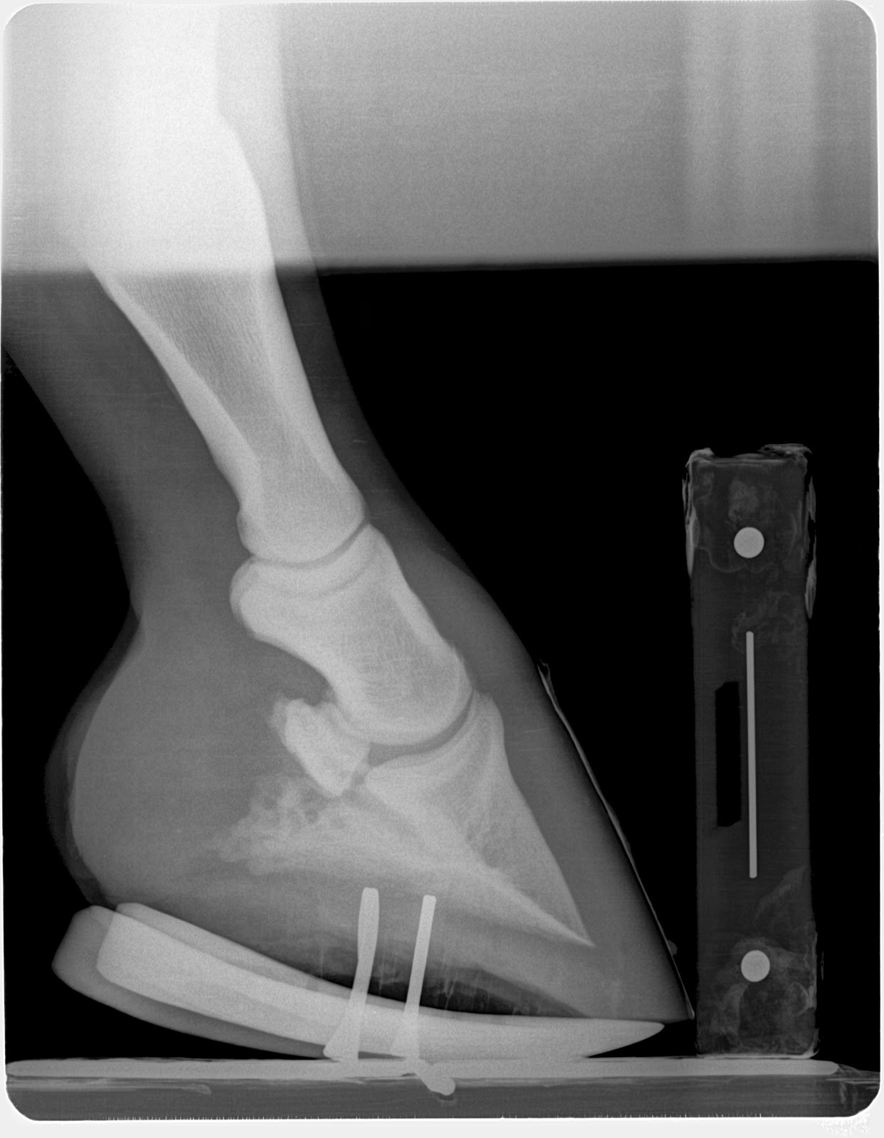
This is a case with navicular bone changes that had responded to increase in palmar angle and reduction in digital breakover but was not consistently going sound. Dr. Redden applied an aluminum rocker rail. Look at the TSA and the distance the navicular is from the proximal p2 between the pre and post shoe radiographs.


This patient has had some undiagnosed recurring lameness. Today no in depth workup was performed but a shoe to enhance foot mass recovery and increase sole depth was applied.


This is a many year chronic laminitis. Goal with the rockered aluminum rail is to decrease DDFT tension unloading the apex of the coffin bone and the tension forces at the horn/lamellar zone. This places breakover in the center of articulation. This will improved compromised circulation in the dorsal region of hoof and a more even hoof growth from toe to heel is expected as well as improved sole depth.


This case had an acute bout of laminitis about 6 months ago. Venograms show that circumflex is above the apex of coffin bone and compressed tightly to tip of coffin bone as well. Dorsal lamellar zone on the right front is broken and a void of contrast is present at coronary plexus. No solar papillae are evident even at an increased to 20 degree palmar angle which should unload DDFT by 60 percent. Treatment included derotational shoeing followed by a deep digital flexor tenotomy.


November 17th reset images from the in depth equine podiatry lecture and demonstration with Dr. Ric Redden, DVM
Below is several follow up images and short discussion of each case. Also look back at the previous blog entry for initial images and therapeutic shoe applied. October clinic images link
White line disease Case: Sole depth improved by 4mm but white line lesion failed to grow down at same rate and decision was made to remove hoof wall to expose oxygen and allow cleaning. Owner reports that he is running around like a youngster again and is more comfy than is has been in a long time.
 |
| 6wks post intial rocker rail note 4mm increase in sole depth in a horse that hasn't grown any sole in years. |
 |
| Reset image |
 |
| First image Oct 6 pre shoe |
 |
| Hoof wall resection to allow cleaning and oxygen to penetrate |


Club foot case: This horse lost the rocker rail shoe applie to the foot opposite the grade 3 club (which is also a club) and regular farrier applied a flat steel keg shoe to keep foot protected. Note the horn lamellar zone divergence. One could call this rotation which would be non specific. The divergence is created by the club syndrome stretching to lower horn to bone attachments. This is confirmed by evaluating the dermal-epidermal junction and measuring the horn zone compared to the lamellar zone. If the lamellar zone was larger than the horn zone one could conclude a laminitis as this is lamellar swelling. In this case it is chronic stretching of the lamellar bone secondary to the constant pull of the deep digital flexor unit.
The Grade 3 club grew more sole in the rocker rail than did the lower grade club in a flat shoe. This information tells us that placing the tendon sling in freedom with the rocker shoe allows better nutrient and blood circulation through unloading of the sole via reduced deep flexor tension. We placed the grade 2 club (Left Front) in a rockered trim with rockered steel keg shoe to also place the tendon sling in release. We will be to see a more rapid sole mass recovery in this hoof as well at the next reset. Owner reports excellent comfort and has adjusted very well to the new shoeing approach.
 |
| Pre shoe radiograph Oct 5 |
 |
| Left front shoe that regular farrier had replaced with flat keg shoe for protection |
 |
| Rockered keg shoe |
 |
| 6 wks post rocker rail application additional 4mm of sole and cup starting to form. All this due to unloading of the deep flexor pull |
Chronic Lamintis case: Farrier was a student and he reports horse is moving very nice. Horse was able to stand comfortably for each shoe reset. Turning and moving very nicely.
 |
| Pre Rocker shoe oct 6 |
 |
| 6 weeks post rocker rail with addition of 4mm of sole and less bulge of sole at apex of frog. |
 |
| Oct 6th pre rocker |
 |
| Left front 6wks post rocker rail. Rocker shoe was removed prior to getting a radiograph. Added 5mm of sole |
 |
| Nov 17th reset with rocker rail. |
 |
| Post nov 17th reset rocker rail. |
Navicular case: Owner reports she was able to work a pattern for the first time in 2 years. The Owners farrier was present and we helped him reset the rocker rails. We plan to maintian the higher palmar angle for the next shoe cycle then began to lower the mechanics/palmar angle. I expect to achieve similar comfort with lower mechanics as the horse remained comfortable even with losing a few degrees of palmar angle secondary to growth. The history is very important here. If horse became more lame at the end of the cycle as the palmar angle decreased, this tells us the hot spot becomes loaded at the lower palmar angle and may require a longer period of higher mechanics.
 |
| RF pre reset on nov17th |
 |
| Post shoe nov 17th |
 |
| Post shoe nov 17th |
 |
| Pre shoe reset on nov 17th |
6 month chronic laminitis case: Owner reports horse is very comfortable, has a much better appetite and very willing to move freely. This case demonstrates the importance the deep digital flexor tendon force applied to a failed lamellar bone. With the loss of the lamellar suspension of the coffin bone, it is allowed to compress the sole at the apex of coffin. No blood, No growth and recurrent abscessation as has occurred in this case. The fragile rim of the coffin bone becomes loses its blood supply and acts like a foreign body. I haven't been able to achieve this level of success with any other approach. Doubling sole depth from 10mm to 20 mm in a matter of 6 wks in chronic laminitis is astonishing.
I do not recommend a tenotomy for every laminitis case and only do so if the venogram shows the circumflex artery at or above the level of the tip of the coffin bone as described by Dr. Ric Redden. However I do recommend considering the forces applied by the ddft to the coffin bone and often use "mechanics" (rockering/wedging) to lesson the tension on a failing system to aid in re-establishing vascular supply.
 |
| Immediately post derotation and deep flexor tenotomy oct 6 |
 |
| Note the rapid growth of sole at dorsal portion of hoof and loss of palmar angle. addition of 10mm of sole |
 |
| Post reset to re establish a zero palmar angle with the shoe. This is necessary to prevent over correction resulting in a negative palmar angle |
 |
| Immediately post derotation and deep flexor tenotomy on oct 6 |
 |
| 6wks post derotational shoeing and deep flexor tenotomy. No reset required as even sole growth is occuring and resetting the shoe does not add any benefits mechanically. |